
Series Title: Structure-Function
A couple of friendly reminders:
- This is not medical advice. The content of this Newsletter is informational only.
- LINKS <-- Yellow highlighted text are helpful links for you to click on.
- You can find much more therapy treatment and intervention content in the Beyond Occupation Blog
- Member only content is at the bottom of the newsletter where you will receive...
- relevent links to resources and/or printables from the BOT Google Drive. If you are a member and have not been added to the drive for content, please request access by clicking this link.
- To go directly to the brand new structure-function folder in the BOT Google Drive for this series, click this structure-function link.
- Clinical conversation starters that take you a bit deeper than the documentation tips in this newsletter.
- If you want to learn more about the Functional Cognition Lab inside the BOT Portal, click here.
Picture This:
Scenario One
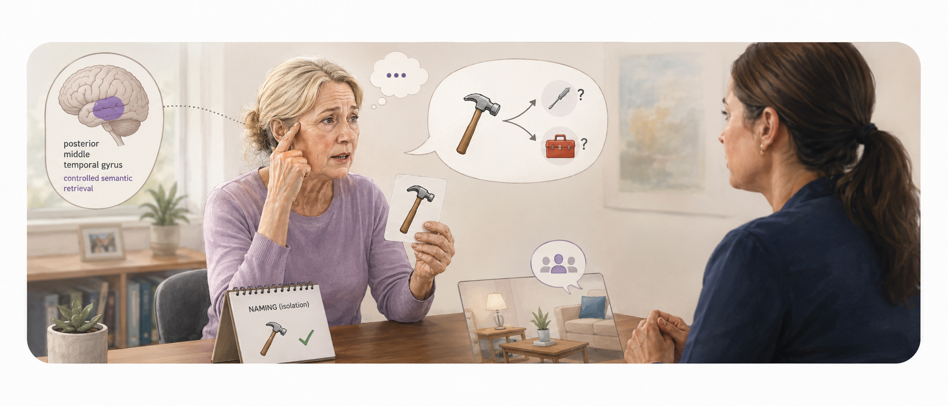
She had a left hemisphere stroke six weeks ago. Her language is not gone. She can speak. She can follow simple instructions. She can name common objects when shown pictures in isolation.
But put her in a real conversation and something breaks down.......
She can't retrieve the right word when the context is too complex. She reaches for a word, gets close, and lands on something related but wrong. She can't find the specific word the situation requires, but she can name the general category it belongs to. This is where some practitioners may stop thinking 'atleast she can speak around the word'. But, I urge you to keep going!
She cannot follow the thread of a story that requires her to hold multiple meanings simultaneously. She understands individual words but loses the narrative. When asked to explain how to use a tool she knows well, she can describe its appearance but cannot retrieve the motoric and semantic details that would tell her hands what to do with it. (See the Multi-Tasks Resourge Page)
The team has documented mild expressive aphasia and word retrieval deficits. The plan includes naming drills and repetition practice.
The naming drills are producing marginal gains in isolation (so, your treatment is technically working....). But, nothing is carrying over to a functional conversation. This is also the kind of patient who may score adequately during structured testing while remaining confused during everyday activity. See Scores Well but Confused for more on that clinical mismatch.
See, we knew it was a language deficit. But, we are at a point in rehab science when precision is necessary. We need to go beyond the "language system" and start asking (1) which part of the language system and (2) how that part may impact other aspects of functional performance. Nobody has asked which part of the language system is failing and so the more precise target goes overlooked: the controlled retrieval of context-appropriate meaning under semantic demand which requires a different intervention than naming drills.
The posterior middle temporal gyrus works alongside executive prefrontal regions to selectively retrieve non-dominant semantic associations and shape conceptual retrieval to fit specific contextual goals (Whitney et al., 2010). When this system is disrupted, your patient can retrieve the most common association for a word but cannot access the meaning the current context requires. They know what a hammer is. They cannot retrieve the specific semantic details that tell them how to use it when the task demands it. Word Repetition practice does not rebuild that system. Controlled semantic retrieval training does.
Scenario Two
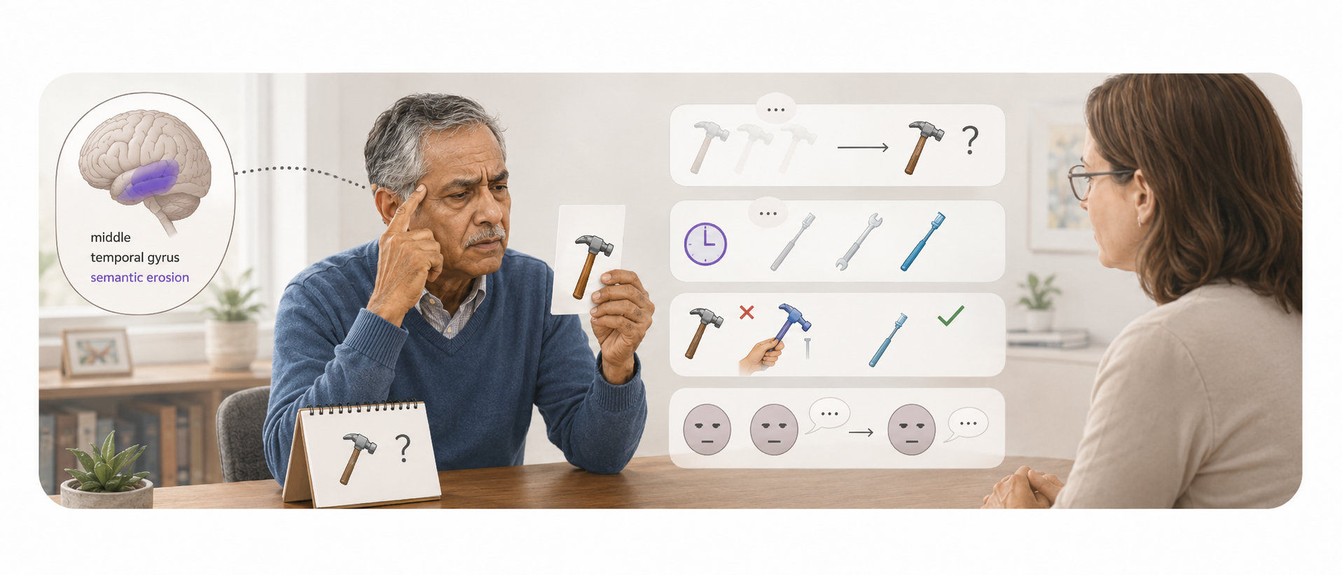
He is sixty-eight years old. Alzheimer's disease, diagnosed fourteen months ago. He is in outpatient cognitive rehabilitation twice per week.
His team is tracking his decline carefully. Naming errors increasing. Category fluency dropping. Object recognition slower. Tool use becoming inconsistent. Social interactions increasingly flat and literall. All of these are accurate, and important observations, but they aren't demonstrating the most precise part. By knowing the structure, you know how all of these symptoms are connected.
They are not connecting progressive naming failure, tool use breakdown, and social communication erosion to the region where all three of those functions live together.
In Alzheimer's disease, plaque-associated microglia in layers three, five, and six of the middle temporal gyrus inversely correlate with memory scores, independent of total amyloid plaque load (Escarião et al., 2026). Early downregulation of excitatory neuronal, synaptic, and mitochondrial pathways occurs in the middle temporal gyrus before widespread cortical involvement (Piras et al., 2025). Reduced intracortical myelin integrity in the right middle temporal gyrus strongly correlates with visuospatial copying impairments in Alzheimer's patients (Ohnishi et al., 2024).
The semantic erosion pattern your team is observing is not generic cognitive decline. It is middle temporal gyrus cortical thinning with a specific layer-by-layer histopathological signature. And knowing that changes what you measure, what you target, and how you interpret rate of change across your plan of care.
What is actually happening:
Where the Middle Temporal Gyrus Is Located
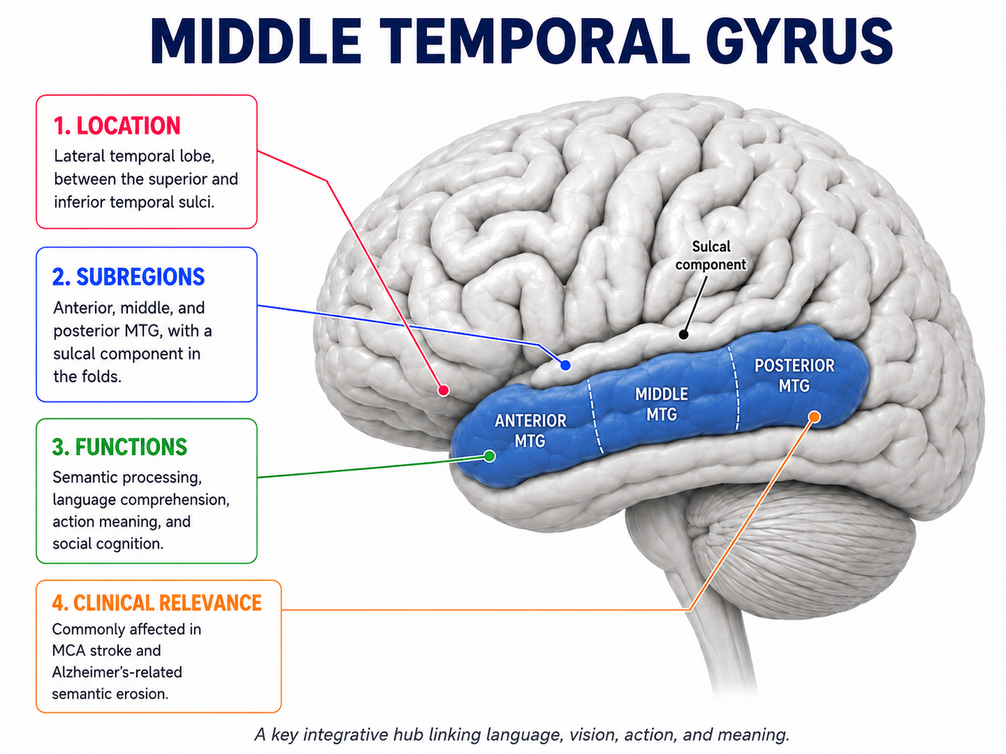
- The middle temporal gyrus is a long fold of cortex located on the lateral surface of the temporal lobe
- It is bordered above by the superior temporal sulcus and below by the inferior temporal sulcus
- It extends from the temporal pole at the front of the brain to the temporo-parieto-occipital junction at the back
- Its main body includes Brodmann area 21, while its posterior portion extends into Brodmann area 37 near the occipital lobe
- The middle temporal gyrus receives most of its blood supply from the middle temporal branches of the middle cerebral artery
- Because of this vascular supply, middle cerebral artery strokes are among the most common causes of middle temporal gyrus disruption
The Middle Temporal Gyrus Is Not One Uniform Region
- The middle temporal gyrus should not be viewed as a single functional area.
- Resting-state functional connectivity and diffusion tractography divide it into four subregions that are arranged along an anterior-to-posterior axis: anterior, middle, posterior, and sulcal regions (Xu et al., 2019).
- Each subregion has its own functional role and pattern of neural connections (Xu et al., 2019).
- Treating all middle temporal gyrus injuries as the same is similar to treating “stroke” as the full diagnosis without considering the specific lesion location.
Anterior Middle Temporal Gyrus
- The anterior middle temporal gyrus acts as a multimodal semantic interface (Davey et al., 2016).
- It combines auditory, visual, and spatial information so that a person can understand words, narratives, and concepts (Davey et al., 2016).
- It contributes to word comprehension, narrative understanding, and retrieval of conceptual memories (Davey et al., 2016).
- This region responds more strongly to spoken words than to visual gestures (Papeo et al., 2019).
- It connects with the anterior temporal lobe, posterior cingulate cortex, and medial prefrontal cortex through the resting-state semantic network (Wei et al., 2012).
Posterior Middle Temporal Gyrus
- The posterior middle temporal gyrus includes the middle temporal visual area, also called V5 or MT
- V5/MT processes the direction, speed, and visual coherence of moving stimuli and contributes to the perception of biological motion, such as recognizing how another person is moving
- The posterior middle temporal gyrus responds more strongly to visual gestures and pantomimes than to spoken words (Papeo et al., 2019).
- It works with executive regions of the prefrontal cortex to retrieve less obvious or non-dominant semantic associations (Whitney et al., 2010).
- Ventral-posterior portions represent object graspability, physical action constraints, and motor details observed during another person’s actions (Xu et al., 2019).
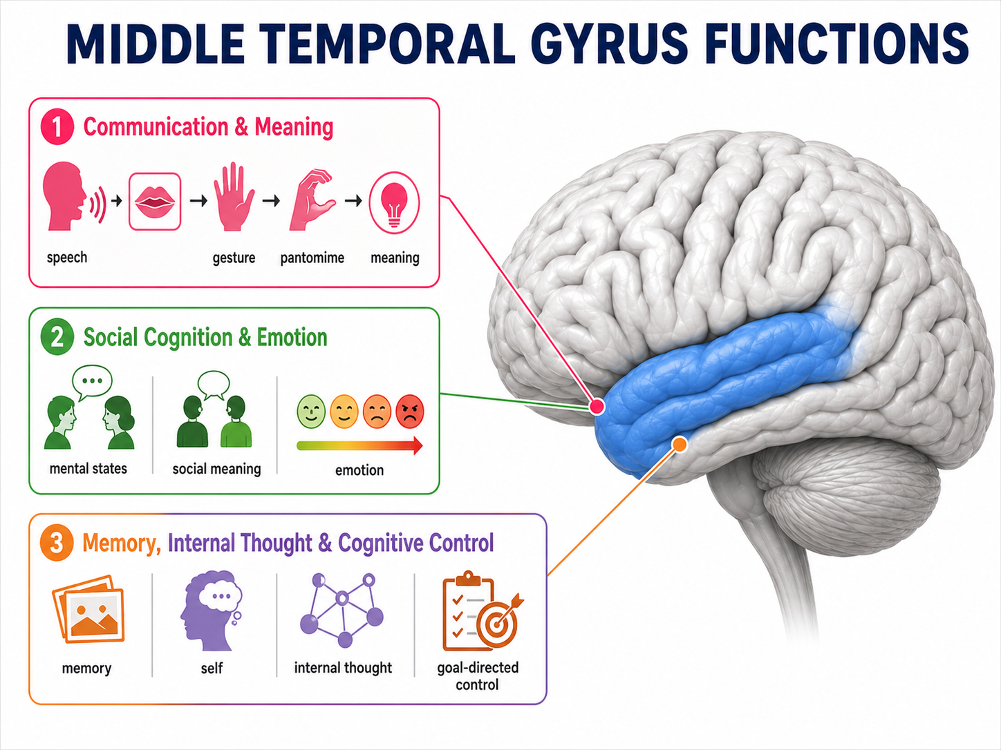
Communication and Meaning
- The middle temporal gyrus helps connect communicative signals with their conceptual meanings (Papeo et al., 2019).
- These signals include spoken words, symbolic gestures, and manual pantomimes (Papeo et al., 2019).
- It helps the brain understand that a sound, movement, gesture, or symbol refers to a specific idea or action.
- It integrates auditory-verbal information with visual-perceptual information (Petrides, 2023).
- This includes matching visible lip movements with speech sounds during the McGurk effect (Petrides, 2023).
Social Cognition and Emotion
- The middle temporal gyrus contributes to Theory of Mind, or the ability to infer another person’s thoughts, intentions, and mental state (Xu et al., 2019).
- It also helps interpret social interactions and the meaning of other people’s behavior (Xu et al., 2019).
- The right middle temporal gyrus is a central network node involved in distinguishing low-arousal emotional stimuli from high-arousal emotional stimuli (Pan et al., 2025).
Memory, Internal Thought, and Cognitive Control
- The middle temporal gyrus is an important node within the Default Mode Network (Briggs et al., 2021).
- Through this network, it contributes to internally generated thought, episodic memory consolidation, and self-referential mental activity (Briggs et al., 2021).
- It is positioned at the structural and functional intersection of the Default Mode Network and the Multiple-Demand Network (Davey et al., 2016).
- The Multiple-Demand Network supports goal-directed cognitive control, flexible thinking, and task management (Davey et al., 2016).
- This position allows the middle temporal gyrus to connect stored knowledge and internally generated information with active task demands.
Why This Region Matters in Rehabilitation
- Damage to the middle temporal gyrus may affect much more than isolated language or visual skills.
- A person may have difficulty combining language, vision, action, memory, emotion, and social information into a meaningful whole.
- The clinical presentation will depend heavily on whether the anterior, posterior, middle, or sulcal portion is affected.
- Therapists should consider whether an apparent memory, language, visual-perceptual, praxis, emotional, or social-cognitive deficit may involve disrupted integration across systems.
- The middle temporal gyrus helps language, vision, action, memory, and social information become meaningful in combination.
Take Action
Member Resource: Medial Temporal Probing Checklist
Four observable clinical signs that the middle temporal gyrus is the disrupted system:
1. Context-specific word retrieval failure with preserved basic naming.
The patient names common objects accurately in isolation but fails when the retrieval demand is contextually specific. They can name a hammer but cannot retrieve the word for the specific type of hammer the task requires. They can name a familiar person but cannot retrieve the semantic details that distinguish that person from a similar one. We may be tempted to say this is a phonological retrieval failure, but it is actually more indicative of a posterior middle temporal gyrus controlled semantic retrieval failure (aka the inability to access non-dominant semantic associations under contextual demand (Whitney et al., 2010). Naming drills may not truly address it. Context-specific semantic retrieval training may help (Stampacchia, S.,et al, 2022).
*This is not just semantic knowledge training. The person may still know what the item is. The impairment may involve semantic control: efficiently retrieving the particular part of that knowledge that matters right now while suppressing stronger but irrelevant associations.
Member Resource: Context-Specific Semantic Retrieval Exercise
2. Tool use and object manipulation breakdown inconsistent with motor capacity.
The patient can describe what an object is and can physically execute the required movements, but the two do not connect into purposeful tool use. They pick up a toothbrush and hold it correctly but cannot generate the motoric semantic program that tells the hand what to do next. Ventro-posterior middle temporal gyrus subregions code object graspability, physical action constraints, and motoric details during action observation (Xu et al., 2019; Davey et al., 2016). When this system is disrupted, the semantic knowledge that should drive tool use is inaccessible at the point of action. This is a middle temporal gyrus apraxia finding, not a motor planning failure in the conventional sense.
3. Visual motion perception failure in dynamic environments.
The patient navigates well in static environments but becomes disoriented, unsafe, or visually overwhelmed in environments with movement like crowded hallways, busy streets, or moving objects in the peripheral field.
In severe cases, moving objects appear as a series of static discontinuous frames (a condition called akinetopsia, produced by bilateral disruption of area V5/MT in the posterior middle temporal gyrus). Unfortunately, rehabilitation may interpret and document this as a "balance problem". It's not. In the world of precision rehabilitation, we label it for the structure-function relationship. It is not a balance problem, it is a visual motion perception problem. It will not respond to vestibular or proprioceptive balance training. It requires environmental adaptation and visual motion-specific intervention.
4. Progressive semantic erosion across naming, category fluency, and social communication simultaneously.
The patient is losing naming accuracy, category fluency, and social communication quality together, across the same time window. When all three deteriorate in parallel, the common denominator is the multimodal semantic interface where all three functions converge. Have you guessed? That's right! The middle temporal gyrus (Davey et al., 2016; Escarião et al., 2026).
In Alzheimer's disease, this pattern reflects layer-specific cortical thinning and plaque-associated microglial accumulation in layers three, five, and six (Escarião et al., 2026). Track the three functions together as a middle temporal gyrus functional composite. Rate of decline across the composite is more clinically meaningful than any single measure in isolation.
The Setup:
Before you choose your intervention, probe the middle temporal gyrus system specifically.
Give the patient a controlled semantic retrieval task. Name an object and ask them to tell you what it is used for in a specific context (not the standard use, but a contextually modified one). For example, ask what a hammer is used for when you are trying to open a paint can with no screwdriver available. Listen for whether they can access the non-dominant semantic association the context requires or whether they default to the dominant meaning regardless of context (Whitney et al., 2010).
Give them a brief tool use observation task. Show them a familiar tool and ask them to demonstrate its use without verbal instruction. Watch whether the semantic knowledge drives the action or whether the action is initiated without the semantic program that should be guiding it (Xu et al., 2019).
Ask them to watch a brief video clip with movement and describe what is happening. Note whether motion perception is intact, whether they can track moving objects, describe direction and speed, and integrate biological motion into a coherent social scene.
Ask them to describe a social scenario (maybe you watch a short youtube video) and note whether they can retrieve the emotional valence, infer the intentions of the people involved, and access the social semantic details that give the interaction its meaning (Pan et al., 2025; Xu et al., 2019).

How to Treat This (Real Sessions)
Additional cognition-based treatment ideas are available in the Cognitive Interventions Dashboard.
Controlled semantic retrieval training:
Naming drills target phonological access to dominant semantic associations. They do not target the posterior middle temporal gyrus controlled retrieval network that accesses non-dominant, context-specific meanings (Whitney et al., 2010).
For patients with semantic aphasia, intervention needs to move beyond naming drills into context-specific retrieval tasks (presenting the patient with a specific situational demand and requiring them to retrieve the semantically appropriate word or concept for that context rather than the dominant association).
Non-Invasive Brain Stimulation moment: Repetitive transcranial magnetic stimulation applied over the left posterior middle temporal gyrus selectively modulates executive semantic control and has been used to investigate the neural mechanisms of language retrieval. Where rTMS is available, left posterior middle temporal gyrus targeting is supported for semantic control deficits.
Controlled semantic retrieval should ultimately be practiced within real tasks. The activities in 12 Functional Cognition Activities for Everyday Tasks in Occupational Therapy can be adapted so the patient must select the meaning, word, object, or action that fits the situation.
Because context-specific retrieval also requires inhibition, working memory, and cognitive flexibility, some patients may benefit from combining semantic retrieval tasks with the activities in 9 Executive Functioning Activities for Occupational Therapy.
Multimodal cueing strategies that incorporate visual imagery, pictures, and gestural cues alongside verbal stimuli engage the middle temporal gyrus's cross-modal semantic interface and improve language comprehension beyond what unimodal verbal stimulation produces. The middle temporal gyrus integrates auditory and visual inputs simultaneously, your treatment should do the same. A word presented with a simultaneous gesture, image, and spoken label recruits more of the middle temporal gyrus semantic network than a word presented alone.
Non-verbal communication retraining using conventional emblems and gestural strategies can bypass verbal retrieval deficits by accessing intact intermediate and posterior middle temporal gyrus gesture pathways (Papeo et al., 2019). The posterior middle temporal gyrus responds more strongly to visual gestures than to spoken words, if the verbal retrieval pathway is disrupted, the gestural pathway may be partially available as a compensatory route. Document which communication modality is producing the best retrieval performance and design the home program around the intact pathway while rehabilitating the disrupted one in session.
Tool use and apraxia retraining:
Interventions for retraining functional object use must engage the ventro-posterior middle temporal gyrus, which integrates motoric gesture details with stored object semantics (Davey et al., 2016; Xu et al., 2019).
This means the intervention cannot separate the object from its semantic context. Practicing the physical movement of toothbrushing in isolation does not engage the middle temporal gyrus semantic program that should be driving the movement. The semantic context (that is, the meaning of the tool, the goal of the action, and the situational demand) must be present during the practice for the middle temporal gyrus network to activate.
For additional occupation-based ways to practice object selection, retrieval, and purposeful use, see 8 Functional Object Retrieval Activities for Occupational Therapy.
Visuospatial constructive rehabilitation exercises including complex figure copying directly address visuospatial construction deficits linked to demyelination and structural atrophy in the right middle temporal gyrus (Ohnishi et al., 2024; Li et al., 2025). Track copying accuracy and visuospatial constructive performance as a middle temporal gyrus functional indicator across your plan of care, particularly in Alzheimer's disease where right middle temporal gyrus myelin integrity predicts visuospatial decline.
Social cognition rehabilitation for patients with anterior middle temporal gyrus disruption may target the networks responsible for inferring intentions and processing social cues (Xu et al., 2019). Emotion discrimination training specifically targeting the right middle temporal gyrus network for distinguishing low-arousal from high-arousal emotional stimuli (Pan et al., 2025) is clinically relevant for patients whose emotional regulation and social participation are affected by middle temporal gyrus disruption.
Environmental adaptations for posterior middle temporal gyrus visual motion failure must account for the fact that the patient is not perceiving motion normally. For patients with akinetopsia or significant visual motion processing impairment, the practitioner may work on on adaptive strategies like static auditory or tactile cues when pouring liquids, crossing streets, or navigating crowded environments that replace the dynamic visual information the posterior middle temporal gyrus can no longer process reliably. Document the visual motion deficit explicitly. It is a safety finding that belongs in every discipline's note and in the discharge plan.
Visual motion and gait navigation:
Practioners addressing balance, visual tracking, and gait navigation in patients with posterior middle temporal gyrus disruption must account for the motion perception deficit as the primary mechanism (Ohnishi et al., 2024). Standard balance training that does not address visual motion processing will plateau. Intervention needs to include controlled visual motion exposure like graded introduction of moving environmental stimuli during functional mobility tasks in order to directly rehabilitate the V5/MT visual motion processing system where neuroplasticity remains available. In dynamic environments like busy hallways or community settings, the patient's safety depends on their ability to track and integrate moving stimuli.
Tracking neurodegenerative functional decline as a middle temporal gyrus composite:
For patients with Alzheimer's disease, therapists across disciplines should monitor progressive semantic memory erosion (examples: naming accuracy, category fluency, object recognition, tool use coherence) as a direct functional indicator of middle temporal gyrus cortical thinning and layer-specific histopathology (Escarião et al., 2026; Li et al., 2025).
Track all three functions together as a composite. When the composite decline accelerates, it may reflect accelerating middle temporal gyrus structural compromise and warrant a referral/follow-up to discuss changing symptoms with neurologist of primary care physician.
Across Systems:
Movement: Your patient's difficulty navigating dynamic environments like busy hallways, crowded community settings, environments with moving objects may not be a vestibular problem or a generalized balance deficit. It may be a posterior middle temporal gyrus visual motion perception problem. Your intervention may need to target motion perception directly with graded visual motion exposure during functional mobility tasks.
Communication: Your patient's word retrieval failure in conversation is not always the same as word retrieval failure on a naming drill. The middle temporal gyrus controlled semantic retrieval network is specifically activated when context demands a non-dominant semantic association (Whitney et al., 2010). If your patient performs adequately on confrontation naming but fails in functional communication, the impairment may not be phonological access. Your treatment may need to target context-specific semantic retrieval, multimodal cueing, and gestural communication as an intact pathway.
Occupation: Your patient's tool use failure may not be a motor planning problem. The semantic program that should be driving the movement is inaccessible at the point of action because the ventro-posterior middle temporal gyrus is not delivering the motoric semantic details that connect object knowledge to purposeful use (Xu et al., 2019). Your intervention may need to embed the semantic context into the practice so that the the tool, the goal, and the situational demand all be present simultaneously for the middle temporal gyrus network to activate. Practicing movements in isolation without semantic context does not engage the system you need to rehabilitate.

Documentation
Brief note on goals: Once the mechanism has been identified, goals should describe the functional retrieval demand rather than simply stating that the patient will “improve word finding.” See Occupational Therapy Goals for Cognition for additional goal-writing examples.
Identify and document:
- Middle temporal gyrus subregion implicated: anterior for multimodal semantic integration and narrative comprehension, posterior for controlled semantic retrieval and visual motion perception, ventro-posterior for tool semantics and action observation, right for emotion discrimination and social arousal processing
- Controlled semantic retrieval probe results: context-specific retrieval task administered; document whether dominant or non-dominant semantic associations were accessible and under what contextual demand failure occurred
- Tool use semantic probe results: object demonstration task administered; document whether semantic knowledge drove action or whether physical execution occurred without semantic program activation
- Visual motion perception status: dynamic environment observation documented; akinetopsia or visual motion integration failure identified as primary safety mechanism where present
- Semantic erosion composite: naming accuracy, category fluency, and object recognition tracked together as middle temporal gyrus functional composite; rate of composite decline documented across sessions
Do not write: patient demonstrates word retrieval deficits and difficulty with tool use. Naming drills and ADL practice initiated.
Write what actually happened:
Controlled semantic retrieval probe administered: patient named common objects accurately in isolation but failed to access contextually specific, non-dominant semantic associations under situational demand. This pattern is consistent with disruption of the posterior middle temporal gyrus–inferior frontal gyrus retrieval network described by Whitney et al. (2010), rather than phonological access failure.
Tool-use semantic probe administered: patient initiated toothbrush use physically but could not generate the motoric semantic program required for purposeful action, indicating ventro-posterior middle temporal gyrus object-semantics failure rather than motor-planning impairment.
Visual-motion observation task administered: patient demonstrated difficulty tracking moving objects in a dynamic video clip and integrating biological motion into a coherent social scene. Posterior middle temporal gyrus V5/MT visual-motion processing impairment was identified as a potential community-safety risk.
The intervention plan targets context-specific controlled semantic retrieval training engaging the posterior middle temporal gyrus–inferior frontal gyrus network, multimodal cueing using simultaneous gestural and visual stimuli, tool-use retraining with full semantic context embedded in practice, and graded visual-motion exposure during functional mobility tasks.

One-Line Clinical Reasoning Starters
Controlled Semantic Retrieval Failure | Posterior MTG-IFG Network Disruption
- Posterior middle temporal gyrus controlled semantic retrieval failure identified as the primary mechanism underlying word retrieval deficits in functional conversation despite adequate performance on confrontation naming tasks
- Patient accesses dominant semantic associations accurately in isolation but fails to retrieve contextually specific non-dominant meanings under situational demand
- Naming drill protocol insufficient to address controlled semantic retrieval failure; context-specific semantic retrieval training targeting the posterior MTG-IFG network indicated as primary intervention
- Context-specific retrieval probe administered and failed; dominant-only semantic access documented as primary middle temporal gyrus outcome measure across sessions
Tool Use Semantic Breakdown | Ventro-Posterior MTG Object Semantics Failure
- Ventro-posterior middle temporal gyrus object semantics failure identified as the primary mechanism underlying tool use breakdown in the presence of intact motor capacity and basic object recognition
- Patient initiates physical execution without the motoric semantic program that should be driving purposeful action, indicating middle temporal gyrus action semantics disruption rather than motor planning impairment
- Tool use retraining must embed the full semantic context to engage the ventro-posterior MTG network; isolated movement practice without semantic context does not activate the disrupted system
- Tool use semantic probe administered and failed; semantic-action disconnection documented as primary finding distinct from apraxia of motor planning origin
Visual Motion Perception Failure | Posterior MTG V5/MT Disruption
- Posterior middle temporal gyrus V5/MT visual motion processing disruption identified as the primary mechanism underlying unsafe navigation in dynamic environments
- Patient demonstrates intact static environment navigation with breakdown in moving environments; deficit reflects impaired direction, speed, and biological motion integration rather than vestibular or proprioceptive failure
- Standard balance and vestibular intervention insufficient; graded visual motion exposure during functional mobility tasks indicated to directly rehabilitate the V5/MT system where neuroplasticity remains available
- Visual motion observation probe administered and failed; dynamic environment safety risk documented as a primary middle temporal gyrus-specific finding requiring environmental adaptation pending system rehabilitation
Akinetopsia | Posterior MTG Bilateral Disruption
- Akinetopsia identified as the mechanism underlying the patient's perception of moving objects as a series of static discontinuous frames, consistent with bilateral posterior middle temporal gyrus V5/MT disruption
- Deficit documented as a neurological sequela of posterior MTG injury, not a visual acuity problem or attentional failure; standard vision screening will not capture this finding
- Environmental adaptations indicated as primary safety intervention: static auditory and tactile cues substituted for dynamic visual information when pouring liquids, crossing streets, and navigating crowded environments
- Akinetopsia documented as a functional safety finding; community discharge planning modified to account for visual motion processing failure in dynamic real-world environments
Multimodal Semantic Integration Failure | Anterior MTG Network Disruption
- Anterior middle temporal gyrus multimodal semantic integration failure identified as a potential mechanism underlying narrative comprehension breakdown and conceptual memory retrieval failure across auditory and visual modalities
- Patient comprehends individual words and isolated stimuli but loses meaning when auditory, visual, and spatial information must be integrated into coherent narrative understanding
- Multimodal cueing strategy indicated: simultaneous gestural, visual, and verbal stimuli engage the anterior MTG cross-modal semantic interface more robustly than unimodal verbal presentation alone
- Narrative comprehension probe administered and failed; cross-modal integration breakdown distinct from phonological processing failure
Social Emotion Discrimination Failure | Right MTG Network Disruption
- Right middle temporal gyrus emotion discrimination network disruption identified as the primary mechanism underlying impaired differentiation between low-arousal and high-arousal emotional stimuli in social contexts
- Patient responds to social interactions with reduced emotional accuracy and flat or mismatched affective responses; deficit documented as a neurological sequela of right MTG disruption, not premorbid personality or mood disorder
- Emotion discrimination training indicated alongside social cognition rehabilitation; emotional regulation strategies alone insufficient without addressing the network-level discrimination failure
- Social arousal discrimination probe administered and failed; right MTG emotional processing deficit documented as primary finding with direct implications for community participation and therapeutic relationship quality
See you in the next newsletter
Author Information:
Michelle Eliason, MS, OTR/L
Occupational Therapist & Functional Cognition Educator
Owner, Buffalo Occupational Therapy
PhD Candidate, Rehabilitation Science
Founder of BOT Portal — a clinical system for real-world cognition
Helpful References:
- Briggs et al. (2021). The unique fiber anatomy of middle temporal gyrus default mode connectivity. Operative Neurosurgery, 21(1), E8–E14.
- Davey et al. (2016). Exploring the role of the posterior middle temporal gyrus in semantic cognition. NeuroImage, 137, 165–177.
- Escarião et al. (2026). Layer-specific colocalization of microglia with amyloid plaques in the middle temporal gyrus predicts cognitive decline in Alzheimer’s disease. Aging and Disease, 17(3), 1568–1589.
- Li et al. (2025). Voxel- and surface-based morphometry in patients with mild-to-moderate Alzheimer’s disease. Frontiers in Aging Neuroscience, 17, 1546977.
- Ohnishi et al. (2024). Associations of demyelination in the right middle temporal gyrus and right precuneus with visuospatial cognitive dysfunction in Alzheimer’s disease. Psychogeriatrics, 25, e13223.
- Pan et al. (2025). Left insula and right middle temporal gyrus dominate cortical network discriminating arousal-dependent emotions. Advanced Science.
- Papeo et al. (2019). The large-scale organization of gestures and words in the middle temporal gyrus. The Journal of Neuroscience, 39(30), 5966–5974.
- Petrides (2023). On the evolution of polysensory superior temporal sulcus and middle temporal gyrus. Journal of Comparative Neurology, 531(18), 1987–1995.
- Stampacchia, S., Hallam, G. P., Thompson, H. E., Nathaniel, U., Lanzoni, L., Smallwood, J., Lambon Ralph, M. A., & Jefferies, E. (2022). Training flexible conceptual retrieval in post-stroke aphasia. Neuropsychological rehabilitation, 32(7), 1429–1455. https://doi.org/10.1080/09602011.2021.1895847
- Wei et al. (2012). Predicting conceptual processing capacity from spontaneous neuronal activity of the left middle temporal gyrus. Journal of Neuroscience, 32(2), 481–489.
- Whitney et al. (2011). The neural organization of semantic control. Cerebral Cortex, 21(5), 1066–1075.
- Xu et al. (2019). Delineating functional segregations of the human middle temporal gyrus with resting-state functional connectivity and coactivation patterns. Human Brain Mapping, 40(17), 5159–5171.
Responses