
Series Title: Structure-Function
A couple of friendly reminders:
- This is not medical advice. The content of this Newsletter is informational only.
- LINKS <-- Yellow highlighted text are helpful links for you to click on.
- You can find much more therapy treatment and intervention content in the Beyond Occupation Blog
- Member only content is at the bottom of the newsletter where you will receive...
- relevent links to resources and/or printables from the BOT Google Drive. If you are a member and have not been added to the drive for content, please request access by clicking this link.
- To go directly to the brand new structure-function folder in the BOT Google Drive for this series, click this structure-function link.
- Clinical conversation starters that take you a bit deeper than the documentation tips in this newsletter.
- If you want to learn more about the Functional Cognition Lab inside the BOT Portal, click here.
Picture This:

Scenario One
He had a small stroke. Left posterior parietal territory. The imaging report said mild infarct. The team said mild stroke. The plan said standard stroke protocol.
Three weeks later he is in outpatient rehab and something is wrong that nobody can name.
He is not weak. His language is intact. He scores adequately on orientation and memory screening. But he cannot tell you what day it feels like. He describes events from last week as though they happened this morning and events from this morning as though they happened years ago. He does not recognize himself in a video recording taken during his last session. He cannot follow the thread of a conversation that requires him to understand what someone else might be thinking or feeling. His wife reports that he seems like a different person. He's not confused exactly, but it seems as if he is just somewhere else.
The team has documented mild cognitive impairment, flat affect, and poor carry-over. The plan has not changed. Nobody is thinking about the precuneus.
The precuneus consumes roughly thirty-five percent more glucose than the average brain region when the brain is at rest (Utevsky et al., 2014; Cavanna, 2007). That extraordinary metabolic demand makes it uniquely vulnerable to indirect ischemic damage and neuroinflammation...not just when the infarct is located there, but whenever vascular disruption compromises the metabolic supply to the surrounding territory (Liu et al).
A stroke located elsewhere can silence the precuneus without touching it. And when the precuneus goes offline, what disappears is not 'memory' in the conventional sense, but the self that organizes memory. The sense of physical ownership of the body. The ability to orient oneself in time. The capacity to imagine what another person is thinking.
That is not mild cognitive impairment. That is precuneus network disruption. And it requires a unique clinical response.
Scenario Two
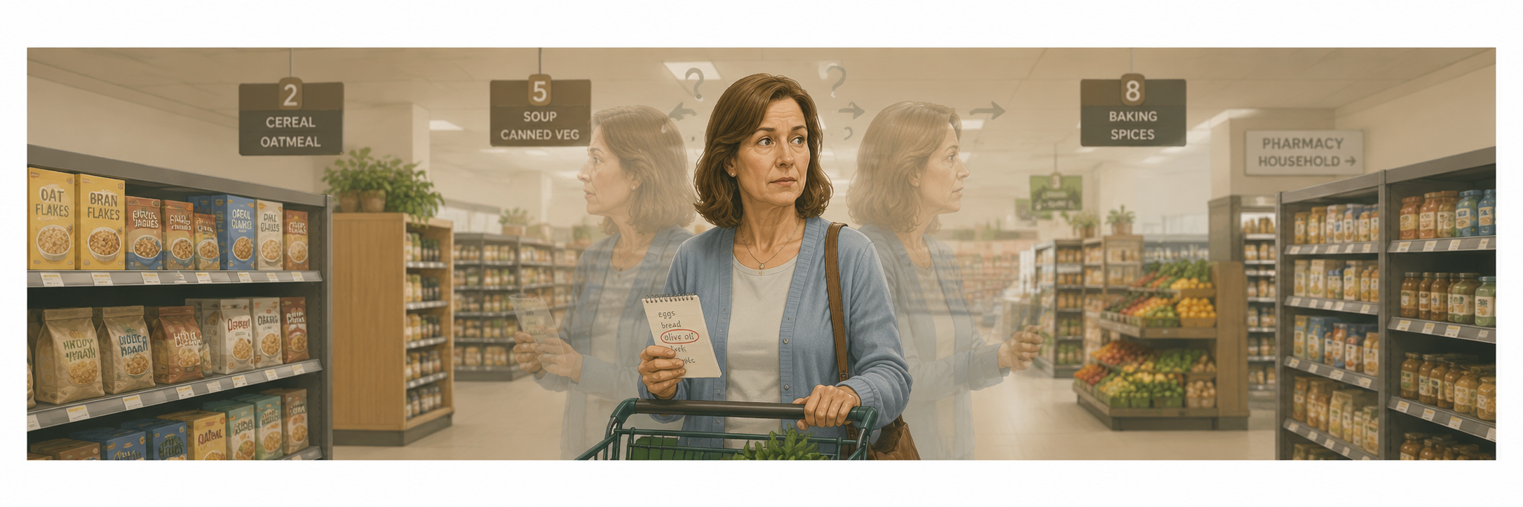
She is fifty-three years old. Early-onset Alzheimer's diagnosis confirmed eight months ago. She is in outpatient cognitive rehabilitation three times per week.
Her program targets memory compensation strategies, medication management, and caregiver education. It is well-designed. It follows the evidence base for Alzheimer's rehabilitation. Unfortunately, it is targeting the wrong system first.
Early-onset Alzheimer's disease specifically and preferentially targets the precuneus before other regions. What deteriorates fastest in early-onset AD is not the semantic memory or language capacity that late-onset AD typically attacks first. It is visuospatial processing. Egocentric movement processing (that is, the ability to orient the self in space relative to the environment). Executive functioning organized around self-referential information. And the visual mental imagery system that allows a person to picture themselves in a future scenario, plan for it, and navigate toward it.
A rehabilitation program that prioritizes verbal memory compensation without first addressing these visuospatial and self-referential deficits is building on a foundation that the precuneus can no longer support. The patient will appear to engage. She will appear to retain. And then nothing will carry over because the system that would make the carryover possible is the one that is failing first.
What is actually happening:
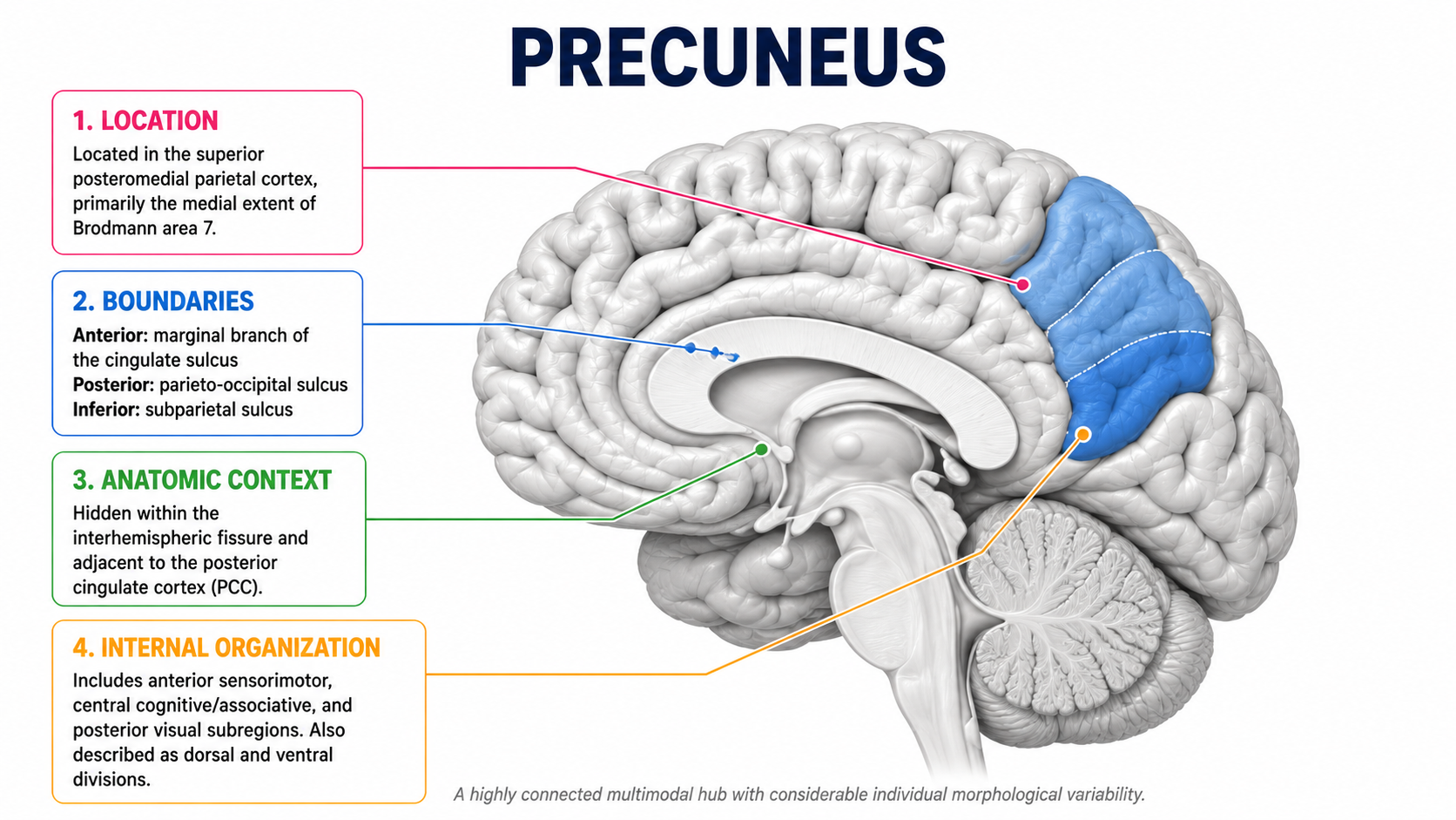
The precuneus sits in the superior posteromedial cortex of the parietal lobe, corresponding primarily to the medial extent of Brodmann area 7 (Messina et al., 2023). It is hidden within the interhemispheric fissure, encased by the sagittal sinus and bridging veins, which is precisely why it was historically difficult to study and why focal lesion research underrepresents its functional significance (Cavanna & Trimble, 2006). It is divided into three functional subregions along an anterior-to-posterior axis: (1) an anterior sensorimotor zone, (2) a central cognitive and associative zone, and a (3) posterior visual zone (Margulies et al., 2009).
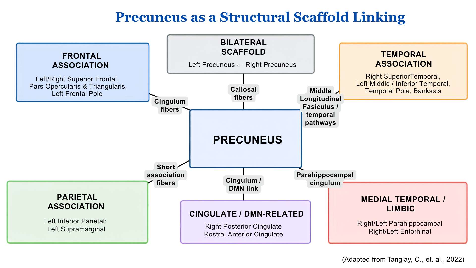
It is the functional core of the Default Mode Network which is the system that activates when the brain is at rest and governs spontaneous cognition, mind-wandering, and internal mentation (Utevsky et al., 2014). It functions as a central topographical node alongside the insula and superior frontal cortex, integrating multimodal networks across the brain (Messina et al., 2023). It lacks direct connections to primary sensory regions, reinforcing its specialized role in elaborating highly integrated associative information rather than processing raw external stimuli (Cavanna & Trimble, 2006).
What does it actually do?
- It decodes 'selfhood' which allows individuals to adopt an embodied first-person perspective during the retrieval of autobiographical memories (the memory happened to them with them in it) (Iriye & St. Jacques, 2025).
- It maintains physical self-awareness and bodily ownership (Stanford Medicine Magazine, 2026).
- It facilitates temporal orientation which is the cognitive ability to distinguish between past, present, and future (Futamura et al., 2025).
- It supports visual mental imagery, with its bilateral activation correlating directly to the vividness of both dynamic and static mental images (Duan et al., 2025).
- It enables 'Theory of Mind' which is the capacity to attribute mental states, empathy, and intention to other people (Messina et al., 2023).
- It engages in complex social language processing, providing the cognitive resources needed to comprehend metaphors, irony, and sarcasm (Valles-Capetillo et al., 2025).
- It selectively deactivates during altered states of consciousness like deep sleep, drug-induced anesthesia, and vegetative states which marks it as on-off switch for self-awareness (Cavanna, 2007).
When it is disrupted by stroke, neurodegeneration, or indirect ischemic damage, what disappears is the integrated sense of self that organizes all other cognitive functions.
That is not a finding you will capture on a standard cognitive screen. It requires clinical observation that is specifically calibrated to look for it.
Take Action
Four observable signs that the precuneus is the disrupted system:
1. Temporal disorientation that does not fit standard confusion.
The patient knows the date when asked but cannot feel the difference between past and future. They describe yesterday's events as ancient history and last year's events as recent. They cannot organize their own timeline. This is not simply 'orientation failure', it is precuneus-mediated temporal orientation failure, or the loss of the felt sense of when things happened relative to now (Futamura et al., 2025).
2. Loss of first-person perspective in autobiographical recall.
The patient can tell you facts about their own life but cannot retrieve them from the inside. They describe personal memories the way they would describe a story they heard (that is, in the third person, detached, with no felt sense of having been there). The precuneus is specifically responsible for allowing individuals to adopt an embodied, first-person visual perspective during autobiographical memory retrieval (Iriye & St. Jacques, 2025). When it is disrupted, the self that inhabits the memory is gone.
3. Impaired Theory of Mind in functional contexts.
The patient cannot accurately infer what another person is thinking, feeling, or intending. They misread social situations. They respond to sarcasm or irony literally. They fail to anticipate how their behavior affects others. This is not personality change or behavioral dysregulation in the psychiatric sense, but is instead precuneus-mediated Theory of Mind failure (Messina et al., 2023). This disruption can and will derail every therapeutic relationship and family interaction until it is named and addressed.
4. Disrupted bodily self-awareness.
The patient does not recognize themselves in video recordings or photographs. They describe their own body in third-person terms. They show reduced awareness of physical sensations that are self-relevant. This reflects the precuneus's central role in maintaining physical self-awareness and bodily ownership (Stanford Medicine Magazine, 2026). Many people document it as flat affect or poor insight, but both of those may put you at risk of missing the root cause of dysfunction.
The Setup:
Before you choose your intervention, probe the precuneus system specifically in your first session.
1. Ask the patient to tell you about something that happened to them five years ago. Listen for whether they remember it and whether they inhabit it In other words, listen for whether they describe it from the inside as something they experienced or from the outside as something they observed.
2. Ask them to tell you what tomorrow will feel like. Can they project themselves into a future scenario and describe it from a first-person perspective? Or does tomorrow feel abstract, flat, and unreachable?
3. Give them a brief social scenario, like a short description of an interaction between two people, and ask them what each person is thinking and feeling. Listen for whether they can generate a mental state attribution or whether they respond only to the literal content of what was said.
4. Ask them to look at a photograph of themselves and describe what they see. Note whether they refer to themselves in the first person or the third.
These four probes tell you whether you are looking at a generic cognitive impairment or a precuneus network disruption. The intervention that follows is different depending on the answer.

How to Treat This (Real Sessions)
Motor imagery:
The precuneus activates heavily during whole-body motor imagery. Motor imagery is imagining complex physical movements like hurdling or navigating an obstacle course activates the precuneus's anterior sensorimotor zone (Ogiso et al., 2000).
For patients who cannot physically execute movements due to injury, illness, or fatigue, motor imagery is a direct precuneus intervention that rebuilds the spatial attributes of movement without requiring physical exertion. Imagery must be first-person and embodied to engage the precuneus in such a way that the patient imagines themselves performing the movement from the inside, not watching themselves from the outside.
Third-person motor imagery does not engage the same system. In tremor-dominant Parkinson's disease, the precuneus drives functional network alterations that contribute to freezing of gait; therapies targeting visuospatial processing through the precuneus can reduce basal ganglia overload and improve gait initiation (Zhang et al., 2025). Deep brain stimulation of the subthalamic nucleus normalizes the structural covariance networks predominantly driven by the precuneus in this population (Zhang et al., 2025).
Temporal anchoring and structured routines
Precuneus atrophy in early-stage Alzheimer's causes severe temporal disorientation causing the patient to lose the felt concept of past versus future, not just the factual knowledge of the date (Futamura et al., 2025).
Compensatory strategies that rely on the patient's ability to orient themselves in time will fail if the temporal orientation system is the thing that is failing. The intervention needs to externalize time. Structured daily routines that anchor the patient to a predictable temporal sequence (that is, same activities, same order, same environmental cues at the same time of day) in order to reduce the demand on a failing internal temporal orientation system.
Cortical thinning of the precuneus after mild stroke is a strong radiographic predictor of Mild Behavioral Impairment, signaling practitioners to preemptively target emotional and behavioral regulation before behavioral symptoms escalate.
Pragmatic language and social communication
The precuneus provides the cognitive resources necessary for understanding sarcasm, irony, metaphors, and socially embedded language (Valles-Capetillo et al., 2025). When this system is disrupted, the patient processes social language literally which means they understand the words but not the social meaning behind them.
Intervention needs to explicitly target these associative networks rather than assuming that intact basic language comprehension means intact pragmatic language comprehension.
For example, in schizophrenia, the core psychotic experience (that is, the inability to harmonize external reality with internal self-perception) stems directly from precuneus network dysfunction (Messina et al., 2023), highlighting a target for cognitive-behavioral and reality-testing approaches in that population.
Resistance training as structural protection:
Engaging in physical resistance training protects the precuneus and hippocampus against structural atrophy and improves white matter integrity in older adults with Mild Cognitive Impairment (Ribeiro et al., 2025). This is not general exercise as wellness. It is a structural intervention targeting the specific regions most vulnerable to MCI-related degeneration.
Mindfulness and attention training:
Six weeks of mindfulness training can measurably increase precuneus grey matter volume (Wikipedia contributors, 2026). Single-session behavioral interventions such as Attention Training Technique can significantly alter regional dynamics and function within the Default Mode Network and precuneus, particularly for individuals with high cognitive-attentional syndrome.
Across Disciplines
PT: Your motor imagery protocol is a precuneus intervention. The specificity of the imagery (first-person and embodied, not third-person and observed) determines whether you are engaging the precuneus or bypassing it. An example would be a patient with tremor-dominant Parkinson's disease or post-stroke motor deficits whose precuneus is compromised needs imagery designed to rebuild the self-in-space representation, not just rehearse the movement sequence.
SLP: Your patient's pragmatic language failure is not a basic language comprehension problem; it is a precuneus associative network failure. When they cannot understand that a colleague's comment was sarcastic, or cannot interpret a metaphor, or cannot follow the social subtext of a conversation, the precuneus is not providing the cognitive scaffolding that social language requires. Your intervention needs to target those associative networks directly, not assume that intact syntax and vocabulary means intact social communication.
OT: Your patient's temporal disorientation, self-referential memory failure, and Theory of Mind impairment are all precuneus findings. They are not generic dementia behaviors or post-stroke personality changes. They are system-specific failures that require system-specific interventions like temporal anchoring, bodily self-awareness training, and structured social cognition tasks embedded in functional ADL contexts.

Documentation
Identify and document:
- Precuneus-specific deficit pattern: temporal disorientation inconsistent with standard orientation failure, first-person perspective loss in autobiographical recall, Theory of Mind impairment in functional contexts, disrupted bodily self-awareness and ownership
- The system implicated: precuneus as the functional core of the Default Mode Network; document distinction from posterior cingulate cortex and from generic cognitive impairment
- Probe results: temporal projection task, autobiographical perspective probe, Theory of Mind social scenario, self-recognition task; document what specifically failed and at what point
- Communicate ulnerability mechanism where applicable: indirect ischemic precuneus damage secondary to high metabolic demand; document that deficits may be present even when the infarct is not located in the precuneus
- Intervention rationale: first-person motor imagery, temporal anchoring, pragmatic language targeting, resistance training as structural intervention, non-invasive brain stimulation where available
Do not write: patient demonstrates mild cognitive impairment, flat affect, and poor carry-over following left posterior parietal stroke. Standard stroke cognitive protocol initiated.
Write what actually happened:
- Patient presents with indirect precuneus network disruption following left posterior parietal ischemic infarct
- Precuneus vulnerability attributed to its exceptionally high resting metabolic demand rendering it susceptible to ischemic compromise beyond the primary infarct territory.
- Precuneus-specific probes administered:
- Temporal projection task failed — patient unable to generate first-person future scenario or organize personal timeline coherently
- Autobiographical memory retrieved in third-person detached perspective without embodied self-referential quality
- Theory of Mind social scenario — patient unable to accurately attribute mental states to described individuals, responding only to literal content
- Self-recognition probe — patient described own image in third-person terms without self-referential acknowledgment.
- Findings consistent with Default Mode Network disruption centered at precuneus rather than standard amnestic or executive impairment profile. Intervention plan targets first-person embodied motor imagery for sensorimotor zone rehabilitation, temporal anchoring through structured daily routine to externalize failed internal temporal orientation system
- Pragmatic language intervention targeting precuneus associative networks for social communication, and resistance training prescribed as structural intervention targeting precuneus and hippocampal volume preservation.
Author Information:
Michelle Eliason, MS, OTR/L
Occupational Therapist & Functional Cognition Educator
Owner, Buffalo Occupational Therapy
PhD Candidate, Rehabilitation Science
Founder of BOT Portal — a clinical system for real-world cognition

Handout 1: Aquired Brain Injury Symptoms Reference
This is your patient and family education resource for understanding what precuneus and parietal lobe disruption looks like in daily life. Direct your patient and family specifically to the Parietal Lobe section of this handout.
Clinical tip: After reviewing the Parietal Lobe section together, add three precuneus-specific symptoms verbally that the handout does not list: the inability to feel where you are in time, the loss of the felt sense of being yourself in your own memories, and the difficulty understanding what other people are thinking or feeling. Ask the family whether they have noticed any of these in addition to the symptoms on the printed list. Their answers will tell you which precuneus functions are most disrupted in daily life and give you your functional intervention priorities. Document which symptoms the family identified at home as your baseline precuneus functional profile.
Helpful Resource 2: SaeboMind Exercises
SaeboMind is a free audio-guided mental imagery program developed by Saebo for patients with upper and lower extremity deficits following stroke or neurological injury. Each episode guides the patient through a first-person embodied mental imagery sequence of a specific functional task like dealing cards, counting coins, opening a medicine bottle, opening doors followed by a brief period of progressive muscle relaxation. Twenty episodes are available and can be accessed on any device through Apple Podcasts, Overcast, or the RSS feed.
Clinical tip: Before assigning any SaeboMind episode as a home program, confirm in session that your patient is generating first-person imagery rather than watching themselves from the outside. A simple in-session check is to ask the patient to close their eyes and imagine reaching for a glass of water. Ask them whether they are seeing their hand reach or feeling their hand reach. Feeling is first-person. Seeing is third-person. If the patient defaults to third-person, coach the shift before sending them home with the audio program. Document which episode was assigned, whether first-person imagery was confirmed in session, and the patient's self-reported vividness rating after the home practice attempt.

One-Line Clinical Reasoning Starters
Indirect Precuneus Disruption | Post-Stroke Metabolic Vulnerability
- Indirect precuneus network disruption identified as the primary mechanism underlying cognitive deficits despite infarct located outside the precuneus territory
- Precuneus vulnerability attributed to its exceptionally high resting metabolic demand, rendering it susceptible to ischemic compromise and neuroinflammation beyond the primary infarct boundary
- Standard stroke cognitive protocol insufficient to address precuneus-specific network failure; skilled intervention targeting Default Mode Network disruption indicated
- Precuneus-specific probes administered to distinguish network disruption from generic post-stroke cognitive impairment; findings documented as primary driver of observed deficits
Temporal Disorientation | Precuneus-Mediated Orientation Failure
- Precuneus-mediated temporal orientation failure identified as the mechanism underlying the patient's inability to organize personal timeline and project into future scenarios
- Deficit inconsistent with standard orientation impairment; patient retains factual date knowledge but has lost the felt sense of temporal position relative to past and future
- Temporal anchoring intervention indicated to externalize the failed internal orientation system through structured daily routines and consistent environmental time cues
- Temporal projection probe administered and failed; inability to generate first-person future scenario documented as primary precuneus outcome measure across sessions
First-Person Perspective Loss | Autobiographical Memory Disruption
- Precuneus disruption identified as the mechanism underlying loss of first-person embodied perspective during autobiographical memory retrieval
- Patient retrieves personal memories in detached third-person narrative without self-referential quality, indicating Default Mode Network failure rather than episodic memory storage deficit
- Skilled intervention targeting self-referential memory encoding and embodied perspective retrieval indicated; compensation strategies relying on intact self-referential processing will not generalize
- Autobiographical perspective probe administered and failed; third-person retrieval pattern documented as primary precuneus-specific finding
Theory of Mind Failure | Pragmatic and Social Cognition Disruption
- Precuneus-mediated Theory of Mind failure identified as the primary mechanism underlying impaired social cognition in functional contexts
- Patient unable to accurately attribute mental states, intentions, or emotional valence to others; responds to literal content of social language without accessing social subtext
- Pragmatic language intervention targeting precuneus associative networks indicated; intact basic language comprehension does not indicate intact social communication capacity
- Theory of Mind social scenario probe administered and failed; literal-only processing documented as primary finding; skilled SLP intervention targeting irony, metaphor, and mental state attribution indicated
Disrupted Bodily Self-Awareness | Physical Self-Perception Failure
- Precuneus disruption identified as the mechanism underlying impaired bodily ownership and physical self-awareness
- Patient describes own body and self-image in third-person terms without self-referential acknowledgment; deficit documented as a neurological sequela of precuneus network failure, not flat affect or poor insight
- First-person embodied motor imagery indicated as direct precuneus sensorimotor zone intervention; third-person imagery does not engage the same system and should not be substituted
- Self-recognition probe administered and failed; third-person self-description documented as primary precuneus-specific outcome measure
Resistance Training as Structural Precuneus Intervention
- Resistance training prescribed as a structural intervention targeting precuneus and hippocampal volume preservation in patient with Mild Cognitive Impairment
- Evidence base supports resistance training as a protection against structural atrophy and white matter integrity loss in the precuneus-hippocampal system specifically
- Resistance training documented as a neuroplasticity and structural preservation intervention, not a general fitness recommendation
- Precuneus and hippocampal volume preservation identified as the primary structural outcome target; intervention intensity and progression documented accordingly
Early-Onset Alzheimer's Disease | Precuneus-First Rehabilitation Priority
- Early-onset Alzheimer's disease identified as a precuneus-first degenerative process with visuospatial and egocentric processing deteriorating faster than semantic or language functions
- Standard late-onset Alzheimer's rehabilitation protocol insufficient; intervention priority restructured to target visuospatial skills, executive functioning, and self-referential processing before verbal memory compensation
- Cognitive rehabilitation program modified to engage visual-spatial and episodic memory jointly, consistent with precuneus-to-hippocampus connectivity mediating synaptic plasticity and executive function outcomes
- Precuneus-targeted non-invasive brain stimulation identified as a supported adjunct intervention in the prodromal and early stages; referral placed where available
See you in the next newsletter
Helpful References:
- Koch, G., Casula, E. P., Bonnì, S., Borghi, I., Assogna, M., Di Lorenzo, F., ... & Martorana, A. (2025). Effects of 52 weeks of precuneus rTMS in Alzheimer’s disease patients: a randomized trial. Alzheimer's Research & Therapy, 17, Article 69. https://doi.org/10.1186/s13195-025-01709-7
- Iriye, H. M., & St. Jacques, P. L. (2025). An embodied perspective: Angular gyrus and precuneus decode selfhood in memories of naturalistic events. Imaging Neuroscience, Article a.93.
- Bruner, E., & Gallareto-Sande, R. (2025). Growth and development of the precuneus in humans from birth to early adulthood. Cerebral Cortex, 35(12), Article bhaf305. https://doi.org/10.1093/cercor/bhaf305
- Jarvers, I., Degmayr, R., Otto, A., Jacob, R., Malloni, W., Kandsperger, S., ... & Brunner, R. (2025). The relationship between precuneus thickness and psychopathology in adolescent anorexia nervosa. International Journal of Eating Disorders. https://doi.org/10.1002/eat.24591
- Zhang, M. X., Zhao, S. Y., Yang, P. D., Wang, H. Z., ... & Meng, F. G. (2025). The precuneus region drives brain network changes in tremor-dominant Parkinson's disease: Insights from brain morphology and clinical variables. MedComm. https://doi.org/10.1002/mco2.70441
- Bruner, E., Gallareto-Sande, R., López-González, F. J., Zhang, L., Sánchez Martín, C., Grothe, M. J., & Sánchez-Juan, P. (2026). Morphological variations and cortical atrophy of the precuneus in normal aging and Alzheimer's disease. Journal of Anatomy, 248, 62-70. https://doi.org/10.1111/joa.70027
- Utevsky, A. V., Smith, D. V., & Huettel, S. A. (2014). Precuneus is a functional core of the default-mode network. The Journal of Neuroscience, 34(3), 932–940. https://doi.org/10.1523/JNEUROSCI.4227-13.2014
- Precuneus atrophy in early-onset Alzheimer's disease: a morphometric structural MRI study. (n.d.). PubMed.
- Karas, G., Scheltens, P., Rombouts, S., van Schijndel, R., Klein, M., Jones, B., ... & Barkhof, F. (2007). Precuneus atrophy in early-onset Alzheimer’s disease: A morphometric structural MRI study. Neuroradiology, 49, 967–976. https://doi.org/10.1007/s00234-007-0269-2
- Song, J., Lu, Q., Zhang, S., He, C., Zhang, T., Yan, H., ... & Shen, Y. (2025). Precuneus-to-hippocampus connectivity links LTP-like plasticity to cognitive function in subjective cognitive decline and mild cognitive impairment. NeuroImage, 324, Article 121636. https://doi.org/10.1016/j.neuroimage.2025.121636
- Valles-Capetillo, E., Giordano, M., & Kana, R. K. (2025). Cognitive resources and the engagement of the precuneus during irony processing. Psychophysiology. https://doi.org/10.1111/psyp.70162
- He, Y., Shrotri, H., Thomson, A. R., Ghassemi-Tari, Z., Puts, N. A., & Chai, X. J. (2025). Regional excitatory-inhibitory balance relates to self-reference effect on recollection via the precuneus: posterior cingulate cortex-medial prefrontal cortex connectivity. The Journal of Neuroscience, 45(25), e2343242025.
- Ribeiro, I. C., Teixeira, C. V. L., de Resende, T. J. R., de Campos, B. M., Silva, G. B., Uchida, M. C., ... & Balthazar, M. L. F. (2025). Resistance training protects the hippocampus and precuneus against atrophy and benefits white matter integrity in older adults with mild cognitive impairment. GeroScience, 47, 5267–5286. https://doi.org/10.1007/s11357-024-01483-8
- Cunningham, S. I., Tomasi, D., & Volkow, N. D. (2017). Structural and functional connectivity of the precuneus and thalamus to the default mode network. Human Brain Mapping, 38(2), 938–956. https://doi.org/10.1002/hbm.23429
- Fletcher, P. C., Frith, C. D., Baker, S. C., Shallice, T., Frackowiak, R. S. J., & Dolan, R. J. (1995). The mind's eye—Precuneus activation in memory-related imagery. NeuroImage, 2(3), 195–200.
- Cavanna, A. E. (2007). The precuneus and consciousness. CNS Spectrums, 12(7), 545–552. https://doi.org/10.1017/S1092852900021295
- Dadario, N. B., & Sughrue, M. E. (2023). The functional role of the precuneus. Brain, 146(9), 3598–3607. https://doi.org/10.1093/brain/awad181
- Zhao, H., Wei, Z., Feng, T., & Feng, P. (2025). The neural substrates responsible for reward sensitivity association with delay discounting. Brain Structure and Function, 230, Article 157.
- Futamura, A., Kinno, R., Hanazuka, Y., Ochi, R., Midorikawa, A., Kitazawa, S., Ono, K., & Kawamura, M. (2025). The precuneus and posterior cingulate gyrus support temporal orientation in Alzheimer’s disease. Brain Communications, 7(6), fcaf424. https://doi.org/10.1093/braincomms/fcaf424
- Cabanis, M., Pyka, M., Mehl, S., Müller, B. W., Loos-Jankowiak, S., Winterer, G., ... & Kircher, T. (2013). The precuneus and the insula in self-attributional processes. Cognitive, Affective, & Behavioral Neuroscience, 13, 330–345. https://doi.org/10.3758/s13415-012-0143-5
- Ogiso, T., Kobayashi, K., & Sugishita, M. (2000). The precuneus in motor imagery: a magnetoencephalographic study. NeuroReport, 11(6), 1345–1349.
- Cavanna, A. E., & Trimble, M. R. (2006). The precuneus: a review of its functional anatomy and behavioural correlates. Brain, 129(3), 564–583. https://doi.org/10.1093/brain/awl004
- Stanford Medicine Magazine. (2026). Tiny brain structure's huge role in a human's sense of physical self. Retrieved from Stanford Medicine.
- Transcranial magnetic stimulation of the precuneus enhances memory and neural activity in prodromal Alzheimer's disease. (n.d.). PubMed.
- Tanglay, O., Young, I. M., Dadario, N. B., Briggs, R. G., Fonseka, R. D., Dhanaraj, V., ... & Sughrue, M. E. (2022). Anatomy and white-matter connections of the precuneus. Brain Imaging and Behavior, 16, 574–586. https://doi.org/10.1007/s11682-021-00529-1
- Borghi, I., Mencarelli, L., Maiella, M., Casula, E. P., Ferraresi, M., Savastano, E., ... & Koch, G. (2025). Dual transcranial electromagnetic stimulation of the precuneus boosts human long-term memory. eLife, 14, Article 104220. https://doi.org/10.7554/eLife.104220
- Duan, S., Li, Q., Yang, J., Yang, Q., Li, E., Liu, Y., ... & Zhao, B. (2025). Precuneus activation correlates with the vividness of dynamic and static imagery: an fMRI study. Frontiers in Human Neuroscience, 19, Article 1516058. https://doi.org/10.3389/fnhum.2025.1516058
- Margulies, D. S., Vincent, J. L., Kelly, C., Lohmann, G., Uddin, L. Q., Biswal, B. B., ... & Petrides, M. (2009). Precuneus shares intrinsic functional architecture in humans and monkeys. Proceedings of the National Academy of Sciences, 106(47), 20069–20074. https://doi.org/10.1073/pnas.0905314106
- Fransson, P., & Marrelec, G. (2008). The precuneus/posterior cingulate cortex plays a pivotal role in the default mode network: Evidence from a partial correlation network analysis. NeuroImage, 42(3), 1178–1184. https://doi.org/10.1016/j.neuroimage.2008.05.059
- Wang, J., et al. (2026). [Article in Scientific Reports - Exact title missing due to source formatting constraints]. Scientific Reports. https://doi.org/10.1038/s41598-026-40813-y
Responses