
Series Title: Structure-Function
A couple of friendly reminders:
- This is not medical advice. The content of this Newsletter is informational only.
- LINKS <-- Yellow highlighted text are helpful links for you to click on.
- You can find much more therapy treatment and intervention content in the Beyond Occupation Blog
- Member only content is at the bottom of the newsletter where you will receive...
- relevent links to resources and/or printables from the BOT Google Drive. If you are a member and have not been added to the drive for content, please request access by clicking this link.
- To go directly to the brand new structure-function folder in the BOT Google Drive for this series, click this structure-function link.
- Clinical conversation starters that take you a bit deeper than the documentation tips in this newsletter.
- If you want to learn more about the Functional Cognition Lab inside the BOT Portal, click here.
Picture This:
Scenario One
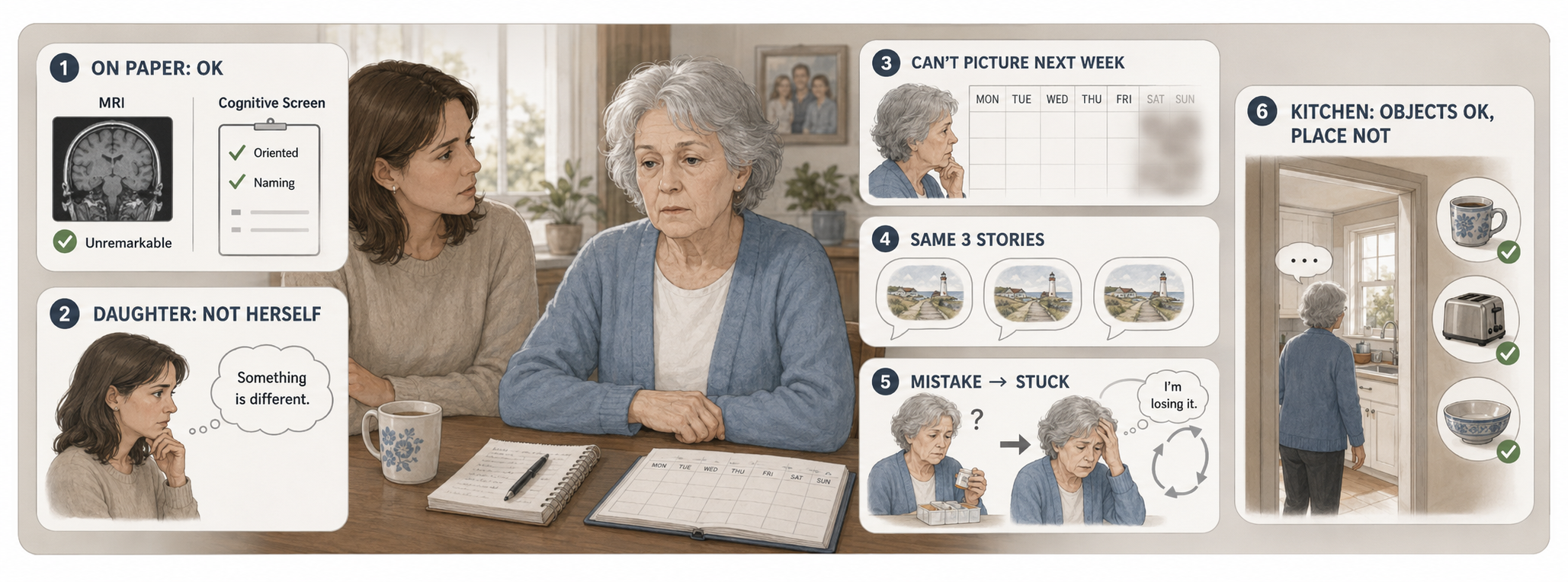
She is seventy-two. Her daughter brought her in because something has changed, and nobody can say what.
Her structural MRI is read as unremarkable for age. She scores adequately on the cognitive screen. Orientation intact. Naming intact. Recall borderline but within normal limits for her education. On paper, there is nothing to treat.
Her daughter says: she is not herself. She sits. She used to plan things, and now she cannot seem to picture next week at all. She tells the same three stories. When she makes a mistake she stops entirely and turns it over and over, saying I'm losing it, I'm losing it, and the rest of the visit is gone. She recognizes every object in her kitchen and still stood in the doorway last Tuesday like she had never been in the room.
The team documented no significant cognitive impairment and discharged her with education and a follow-up in six months. But here's the thing...nobody asked why a normal-looking brain produces that kind of functional presentation.
The posterior cingulate cortex is one of the first regions in the brain to show hypometabolism and tau pathology in aging (Counts et al., 2025). Neurochemical and microstructural decoupling in the PCC has been proposed as an early biomarker that precedes overt cognitive decline (Sun et al., 2025), and MR spectroscopy shows GABA and glutamate imbalance in the PCC in prodromal Alzheimer's disease that may be detectable years before major structural MRI change (Sun et al., 2025). This means that the imaging can be normal and the region can already be failing.
That is not 'no significant cognitive impairment'. That may actually be a metabolic failure in the brain's most metabolically expensive region, presenting exactly the way that failure presents-- planning, self-referential fixation, and in the felt sense of a familiar place. None of these things can be captured by traditional cognitive screens.
Scenario Two
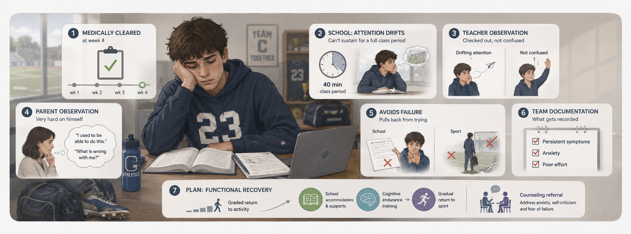
He is seventeen. Sport-related concussion five months ago. Medically cleared at week four.
He has not gone back. Not to the field, not really to school. He says he is fine and then cannot sustain attention through a forty-minute period. His teachers describe him as checked out rather than confused. His parents describe a kid who is now relentlessly hard on himself, constantly saying things like 'I used to be able to do this, what is wrong with me', and who will not attempt anything he might fail at.
The team has documented persistent post-concussive symptoms, anxiety, and poor effort. The plan is graded return-to-activity and a counseling referral. Both of those responses may be correct, but we want to see the situation for what may also be happening because knowing what part of the brain may be breaking down may help guide our decision-making process.
Posterior cingulate dysfunction following traumatic brain injury specifically predicts attentional impairment (Leech & Sharp, 2014). The PCC is used as an MR spectroscopy target in brain injury research precisely because it is a highly metabolically active region thought to play an important role in attention-driven processes, and because it is known to show measurable metabolic change after TBI (Moseley et al., 2026).
And the self-critical loop is not separate from the attention problem. Brooding rumination (that is, the why can't I do this loop) is tied to medial prefrontal–PCC connectivity in depressed adolescents (Duan et al., 2026). PCC activity has been framed as the signature of being caught up in experience, a proposed general mechanism underlying self-referential processing, evaluation, and judgment (Brewer et al., 2013).
He is not putting in poor effort. His attention is lapsing inward and he cannot get back out, and the loop he lands in is the same network that is supposed to be releasing him into the task.
Member Resources:
Ranchos Las Amigas: Grading, Interventions, Questions to Ask
Common Brain Injury Symtoms by Structure Illustration
What is actually happening:
Where it is:
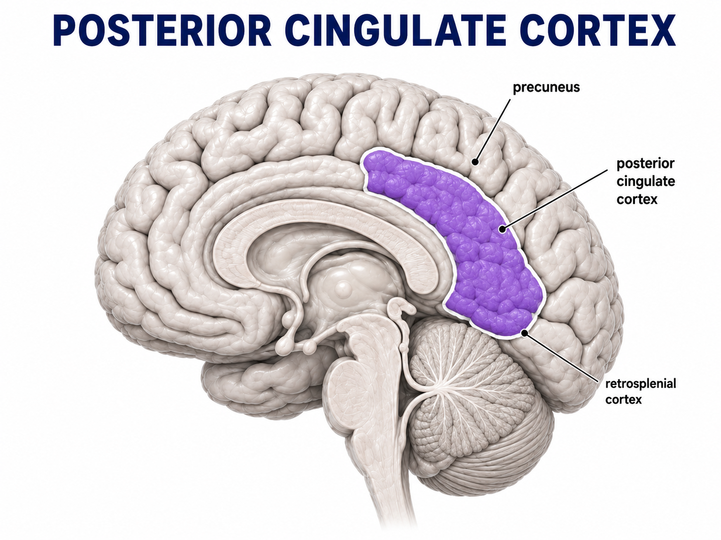
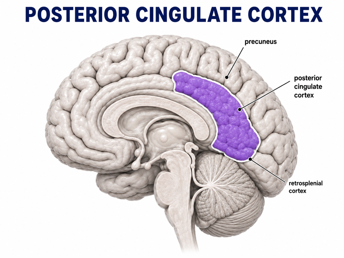
The posterior cingulate cortex (PCC) forms the back portion of the cingulate cortex. It curves around the back of the corpus callosum and sits along the inner surface of the parietal lobe (Foster & Koslov, 2025). The precuneus sits just above it, while the retrosplenial cortex, including Brodmann areas 29 and 30, sits just below it (Foster & Koslov, 2025; Leech & Smallwood, 2019).
Anatomically, the PCC includes Brodmann areas 23 and 31 and can be divided into dorsal and ventral portions (Leech & Sharp, 2014; Leech & Smallwood, 2019). A recently identified groove within the dorsal PCC, called the inframarginal sulcus, appears to mark a transition between the PCC and nearby frontoparietal cognitive-control regions. This area also appears sensitive to cortical thinning associated with aging and Alzheimer's disease (Foster & Koslov, 2025).
The PCC is considered paralimbic cortex, meaning it sits between higher-level association cortex and more traditional limbic structures involved in memory and emotion, such as the hippocampus (Leech & Sharp, 2014).
What it is wired to do:
The dorsal PCC is strongly connected with regions involved in attention and executive control, including the dorsolateral prefrontal cortex, frontal poles, inferior parietal cortex, and superior temporal sulcus. The ventral PCC and nearby retrosplenial cortex have stronger connections with the medial temporal lobe, hippocampus, and ventromedial prefrontal cortex (Leech & Sharp, 2014; Leech & Smallwood, 2019).
The PCC also communicates with the thalamus, striatum, and several other major brain systems. Studies examining the brain's white matter connections consistently identify the PCC as a major hub, meaning it is positioned to exchange information across many different networks (Leech & Smallwood, 2019).
Three broad connection patterns help organize the region: connections within the posteromedial cortex itself, connections between the dorsal PCC and frontoparietal regions, and connections between the ventral PCC and medial temporal memory systems (Foster & Koslov, 2025).
Here is the part that matters clinically: the PCC does not directly connect with primary sensory or motor cortex (Leech & Sharp, 2014; Foster & Koslov, 2025; Leech & Smallwood, 2019).
Instead, it sits near the far end of the brain's processing hierarchy. In other words, it is far removed from basic sensory input and simple motor output. By the time information reaches this system, the brain has already done a great deal of processing around meaning, context, memory, attention, and personal relevance (Leech & Smallwood, 2019; Foster & Koslov, 2025).
So the PCC is not primarily concerned with whether a person can see the cup or move the arm toward it. It is more involved in what that experience means, how it connects with previous experience, where attention goes during the task, and how the person relates the experience back to themselves.
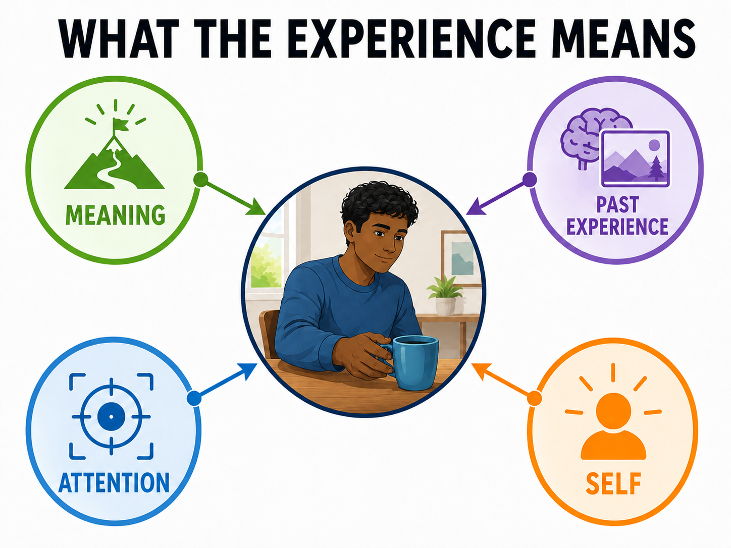
What it does.
The PCC is a major part of the default mode network (DMN), a brain network that becomes especially active during internally focused thinking. This includes autobiographical memory, imagining the future, thinking about oneself, and periods of rest when the mind is not focused on an outside task (Leech & Sharp, 2014).
It acts as a high-level association hub, helping bring together information from executive systems and memory systems so that previous experience can influence what is happening now (Foster & Koslov, 2025). Along with the medial prefrontal cortex, it supports self-referential processing, meaning the way we relate thoughts, memories, emotions, and experiences back to ourselves (Chuda et al., 2026; Joss et al., 2025).
There also appears to be some specialization within the region. The mid-dorsal PCC and retrosplenial cortex contribute to recalling past events, while the posterodorsal PCC is particularly involved in representing personally familiar places (Sugiura et al., 2005).
Interestingly, recognizing a familiar place activates this system differently than recognizing a familiar object. That tells us that even within the PCC, the brain handles spatial familiarity and object familiarity differently (Sugiura et al., 2005).
The PCC has also been linked with spatial orientation, visuomotor coordination, self-referential memory, and evaluating the emotional importance of incoming information (Sun et al., 2025; Dang et al., 2025).
Leech and Smallwood (2019) offer a particularly useful way to think about this clinically. They argue that the PCC may contribute less to one specific cognitive skill and more to how cognition unfolds over time.
Think about memory, navigation, understanding a story, or following your own train of thought. These abilities require the brain to continuously organize information, decide what matters, connect the present with the past, and adjust attention as the situation changes.
There are still several competing theories about exactly what the PCC does. Some focus on its role in internally directed attention and attention lapses. Others emphasize self-generated thought and social cognition. Another view suggests that it helps monitor performance and coordinate activity across large brain networks (Leech & Smallwood, 2019).
Leech and Sharp's Arousal, Balance and Breadth of Attention model makes a similar argument. The dorsal PCC may help regulate how stable or flexible whole-brain activity needs to be at a given moment (Leech & Sharp, 2014).
Research in nonhuman primates also suggests that PCC neurons respond when an individual is deciding whether to keep using a current strategy or explore another option. They may help signal when an approach is no longer useful and when it is time to change course (Leech & Smallwood, 2019).
The self-fusion piece...
Brewer et al. (2013) connect PCC activity with being caught up in an experience. This isn't talking aobout whether someone is thinking about themselves...we all do that! The issue is whether the person can recognize a thought as a thought or whether they become completely absorbed in it.
For example: I am having the thought that I can't do this is different from: I can't do this. In the first statement, there is some distance between the person and the thought. In the second, the thought is experienced almost like a fact.
Across Brewer, the ABBA model, and Lamas-Morales et al. (2025), higher or less-regulated PCC activity appears to be associated with becoming fused with self-referential thoughts, including rumination, self-judgment, and getting pulled deeply into an internal narrative.
Reducing that identification is associated with the ability to step back, notice the thought, and continue functioning without being completely carried away by it. Psychology and contemplative literature often call this decentering or cognitive defusion. That does not mean the PCC is simply a "self-monitoring center." Instead, it appears to relate to the quality of the person's relationship with their own thoughts.
The PCC is some of the most expensive real estate in the brain.
The PCC is one of the most metabolically active regions of the brain when a person is resting (Leech & Sharp, 2014). Its blood flow can be substantially higher than the brain average. Yet when a person turns their attention toward an outside task, PCC activity often decreases (Leech & Smallwood, 2019). Interesting, right???
High energy demand. Major network hub. Early vulnerability in aging and neurodegenerative disease.
Take Action
You cannot observe one of these behaviors and conclude, "the patient has PCC damage." What you can do is recognize a cluster of behaviors that may involve the posterior cingulate/default mode system, especially when the patient's basic cognitive screening does not explain what you are seeing. Then, you can change what you assess, what you track, and how you structure treatment.
1. Self-critical thinking that lasts much longer than the error that triggered it.
The patient makes a normal, expected mistake.
The mistake itself is not the major problem.
The problem is the next ten minutes.
I'm losing it. I used to be able to do this. What is wrong with me?
The patient is no longer simply noticing the thought. They have become absorbed in it.
The task stops. Attention narrows. Performance falls apart. The session struggles to recover.
Getting "caught up" in experience has been linked with PCC-related processes (Brewer et al., 2013), and brooding rumination has been associated with medial prefrontal-PCC connectivity (Duan et al., 2026).
Instead of automatically documenting this as anxiety or poor frustration tolerance, describe exactly what happened and how much it affected function.
2. Attention that disappears inward rather than slowly wearing out.
This looks different from fatigue-related attention failure.
With fatigue or reduced cognitive endurance, you may see performance gradually worsen as the session continues.
This pattern can be sudden.
The patient is completing the task and then seems to disappear mentally for a moment. When their attention returns, they may have no idea how much time passed.
Ask: Where was your attention just now?
Sometimes they can tell you exactly where it went, but often the content is internal and self-related. For example, I was thinking about how I used to do this. I was wondering what my wife thinks of me now. I was thinking about everything I can't do anymore.
PCC dysfunction after TBI has been associated with attentional impairment, and one theory of PCC function connects it with these kinds of attention lapses (Leech & Sharp, 2014; Leech & Smallwood, 2019).
What that means for you is that maybe you document how long someone can sustain attention and track how often attention leaves the task and where it goes.
3. Difficulty letting go of a strategy that has stopped working.
The patient keeps doing the same thing.
The strategy failed three attempts ago, but they are still using it.
This does not always look like the classic perseveration you may associate with frontal dysfunction.
Instead, the patient seems unable to recognize that this is not working anymore. I need another approach.
PCC neurons have been linked with evaluating choices and signaling when it may be time to leave an unsuccessful strategy and try something else (Leech & Smallwood, 2019). Clinically, we can measure how long it takes the patient to abandon the failing strategy and how many attempts occur before they change course giving you something different from simply documenting "impaired cognitive flexibility."
4. Loss of the personal sense of a familiar place, while familiar objects remain recognizable.
This one is subtle.
The patient recognizes the refrigerator.
They recognize the coffee maker.
They recognize the kitchen table.
But standing in their own kitchen somehow does not produce the normal sense of 'I know this place. This is my kitchen. I know what it feels like to be here.'
The posterodorsal PCC appears to contribute specifically to representations of personally familiar places, and this can be separated from the recognition of personally familiar objects (Sugiura et al., 2005).
This is also different from other navigation problems.
Entorhinal dysfunction is more about building and maintaining the spatial map.
Retrosplenial dysfunction is more about using landmarks and direction to orient yourself.
The PCC finding is more about the personal familiarity and self-relevance of the place.
Different problem. Different probe. Potentially different treatment target.
The Setup:
Four probes.
1. Attention lapse probe with content sampling.
During a longer functional task, interrupt the patient at three or four unpredictable points and ask:
Where was your attention just now?
Record how often attention has left the task.
Then record where it went:
- External and related to the task
- External but unrelated to the task
- Internal and self-referential
You are not simply asking whether the patient was distracted.
You are trying to identify the type of distraction.
If self-referential internal thinking repeatedly pulls attention away from the task, that becomes a clinically useful pattern to track.
2. Decentering probe.
Immediately after an error, ask what the patient said to themselves.
Listen carefully to the wording. 'I had the thought that I'm useless' is different from 'I'm useless'.
The first statement shows some distance from the thought. The second is fused. The person is treating the thought as reality. The difference between the first and second target may be trainable (Lamas-Morales et al., 2025).
You can document how many self-critical statements are fused versus distanced across the session. This information may be important for an interdisciplinary team.
3. Strategy disengagement probe.
Create a task where one approach works at first but eventually becomes ineffective.
For example:
- A reaching setup where the original method becomes inefficient
- A sorting or planning activity where the rule changes
- A route where the original path becomes blocked
- An ADL task where the usual strategy no longer works
Measure how many attempts and how much time pass before the patient abandons the ineffective approach.
Do not measure only whether they eventually solve the problem. The finding is how easily they recognize that this strategy is no longer working.
4. Personally familiar place probe.
Ask the patient to describe their own kitchen.
Do not ask for a list of objects initially. Ask: What is it like to stand in your kitchen? Where do you feel like you are in the room? What feels familiar about being there?
Then ask them to name and describe familiar objects from the same environment.
Compare the two.
If familiar objects remain easy to represent but the familiar place itself feels poorly formed or strangely disconnected, you may be seeing the pattern described by Sugiura et al. (2005). If navigation is also a concern, run this alongside the entorhinal route-description probe. Together, these probes may help you separate: spatial map problems, directional/heading problems, and personal-place familiarity problems.

How to Treat This (Real Sessions)
Additional cognition-based treatment ideas are available in the Cognitive Interventions Dashboard.
Disclaimer: There is currently no established PCC-targeted stimulation protocol for rehabilitation. Researchers have proposed that highly individualized brain-network mapping may eventually be needed before targeted neuromodulation of this area is possible because PCC organization varies considerably from person to person (Foster & Koslov, 2025; Leech & Sharp, 2014). Researchers are also studying molecular markers within the PCC, including microRNAs associated with cognitive resilience, MCI, and Alzheimer's disease (Counts et al., 2025). Those are exciting precision-medicine directions, but they are not current rehabilitation interventions that appear in research as they are, but are applications of the research being published on this region.
Decentering and defusion coaching embedded in the functional task.
This is probably the intervention with the clearest connection between the proposed mechanism and something you can actually do during treatment.
And it costs nothing but a change in language.
Cognitive defusion means helping the patient notice a thought as a mental event rather than automatically treating it as a fact. You are not turning the OT or PT session into psychotherapy or meditation. You are changing what happens when an internal thought begins interfering with functional performance.
Instead of immediately reassuring: 'You're doing great.' Try identifying the process: You're having the thought that you're losing it. Can you notice that thought and keep your hand on the cup?
The goal is not to convince the patient that the thought is wrong. The goal is to help them create enough distance from the thought that they can continue participating in the task. Practices involving meta-awareness and cognitive defusion have been associated with reduced rigid self-focused thinking, and reduced identification with thoughts has been associated with reduced PCC activity in experienced practitioners (Lamas-Morales et al., 2025).
When this loop appears, coach it in the moment by tracking: How often does the patient become caught in the loop? and How long does it take them to return to the task? Those are functional outcomes you can follow.
Brief priming before the hard work.
A brief mindfulness-based neurofeedback intervention used before a longer skills-based treatment provides an interesting model for how a short PCC/default-mode-network technique might prepare the patient for later work (Jones et al., 2026). Real-time fMRI neurofeedback can actually teach people to change PCC/DMN activity, but that is a research technology, not something most rehabilitation clinics have available.
The useful clinical idea is the structure: Use a brief attentional or self-regulation activity before the demanding cognitive-motor work rather than saving it as a relaxation activity for the end of the session.
You might document this as attentional priming, much like you would document an aerobic or sensory-motor primer before the main intervention.
Self-judgment as a treatment target with its own plan.
An eight-week Mindful Self-Compassion program produced measurable changes in PCC connectivity along with reductions in self-judgment (Joss et al., 2025). The program included techniques such as compassionate breathing, self-soothing touch, and structured self-compassion practice.
Here's the thing, that does not mean every rehabilitation clinician needs to deliver an eight-week mindfulness program. The clinical point is that self-judgment may itself interfere with function and participation. Instead of treating it as something completely separate from rehabilitation, consider how it is affecting task performance and whether it needs to be addressed directly or through interdisciplinary referral.
For adolescents, rumination-focused CBT that specifically targets brooding may also be a more precise referral option than simply recommending "CBT for anxiety" (Duan et al., 2026).
Recognize that ordinary rehabilitation may already influence these networks.
This is an important one. After total hip arthroplasty, changes in connectivity between the insula and the default mode network/PCC were associated with decreases in both pain and pain catastrophizing (Chuda et al., 2026).
The patients were receiving ordinary postoperative rehabilitation including range of motion, strengthening, and gait training. There was not necessarily a separate psychological intervention producing the effect.
A graded, successful physical experience may influence more than strength or range of motion. It can also change how the person experiences their body, their pain, their ability, and themselves.
That gives you a stronger rationale for thoughtful grading, pacing, repetition, and successful task experience.
You do not need to claim that your strengthening exercise "treated the PCC." You can recognize that functional rehabilitation takes place inside whole brain networks, including systems involved in self-perception and meaning.
Build strategy-switching into the session deliberately.
If one PCC-related function involves recognizing when a strategy is no longer productive, then a session made entirely of predictable tasks using one successful strategy never really challenges that ability (Leech & Smallwood, 2019). Build in moments where the patient has to change course.
Change the rule. Move the object. Block the usual route. Change the sequence. Make the first strategy less efficient. Then track time-to-switch or attempts-before-switching across sessions.
This is similar to perturbation-based treatment used elsewhere in rehabilitation, but here the target is not motor prediction. The target is recognizing: My current approach is not working anymore.
Read the body when the words are not available.
Kinesthetic empathy refers to a clinician's ability to use a patient's movement qualities as information about their experience or state (Vaisvaser, 2026). This may be particularly helpful when a patient cannot clearly describe what they are experiencing.
The markers that are familiar to rehabilitation clinicians include muscle tone, breathing, posture, rhythm, movement effort, and use of space.
Research described by Vaisvaser (2026) suggests that the brain separately processes not only what movement another person is making, but also how that movement feels (its rhythm, force, effort, and energy).
Another useful idea from this literature is the concept of synchronization, desynchronization, and repair. Sometimes clinician and patient fall out of rhythm. The task goes poorly. Frustration rises. Communication becomes less effective. Instead of viewing that moment only as a treatment failure, it can become an opportunity to notice the mismatch, adjust, and reconnect.
Creative arts approaches including dance, movement, art, music, and drama have also been proposed as ways of influencing self-referential and predictive brain networks (Vaisvaser, 2021; Vaisvaser, 2026).
At this point, treat that literature as a rationale for interdisciplinary referral and collaboration, not proof of a specific rehabilitation outcome.

Documentation
Brief note on goals: Once the mechanism has been identified, goals should describe the functional retrieval demand rather than simply stating that the patient will “improve word finding.” See Occupational Therapy Goals for Cognition for additional goal-writing examples.
Identify and document:
- PCC-pattern deficit cluster: attention lapses with self-referential content; self-critical thinking that becomes much larger than the error that triggered it; difficulty abandoning an ineffective strategy; reduced sense of a personally familiar place despite intact familiarity with individual objects.
- System implicated: posterior cingulate cortex as part of the default mode network and as a major association hub. Distinguish it from the precuneus, retrosplenial cortex, and entorhinal cortex when the clinical problem involves navigation or spatial familiarity
- Probe results: number and content of attention lapses; fused versus distanced self-statements after errors; time and number of attempts required to change an ineffective strategy; personally familiar place versus familiar object performance.
- Intervention rationale: cognitive defusion or decentering coaching during functional activity; brief attentional priming before demanding work; directly addressing self-judgment when it interferes with participation; intentionally building strategy changes into functional tasks; graded successful task experiences.
Do not write:
Patient demonstrates mild attention deficits and anxiety, with frequent negative self-statements limiting participation. Encouragement provided.
Write what actually happened:
Patient demonstrates a cognitive-behavioral pattern involving attention regulation, self-referential processing, and strategy monitoring. Attention lapse probe completed during functional standing-tolerance activity. Patient demonstrated four attention lapses over twenty-two minutes. Three of four lapses involved self-referential or evaluative thoughts when sampled. Performance did not progressively decline across the session, suggesting that attention was being intermittently captured by internal content rather than gradually declining because of fatigue.
Decentering probe completed following in-session errors. Patient used fused self-critical statements during five of six opportunities and did not spontaneously use language that created distance from the thought. Mean time required to return to task was six minutes. Self-critical thought episodes interfered with session productivity more than the underlying motor error itself.
Strategy disengagement probe completed during transfer training. Patient continued using an ineffective transfer strategy across seven attempts without independently modifying the approach. Findings suggest difficulty recognizing and disengaging from an unsuccessful strategy. Time and attempts required to change strategies will be tracked across sessions.
Personally familiar place probe completed. Patient accurately named and described familiar household objects but had difficulty describing the first-person experience of standing within her own kitchen. A difference between familiar-object and familiar-place representation was observed. Additional assessment is indicated to distinguish personal-place familiarity from spatial-map or directional-orientation difficulties.
Intervention will target cognitive defusion during functional task performance, brief attentional priming before demanding cognitive-motor activity, deliberate strategy changes within ADL tasks with time-to-switch tracked as an outcome, and graded successful task experiences. Functional impairment despite relatively unremarkable structural imaging will continue to be documented as part of the rationale for skilled intervention.

One-Line Clinical Reasoning Starters
Self-Referential Fusion | PCC-Mediated Session Interference
Fusion with self-referential evaluative content identified as the primary rate-limiting factor for session productivity, exceeding the functional cost of the underlying motor impairment Patient produces fused rather than decentered self-statements following in-session error, with prolonged recovery latency before task re-engagement Cognitive defusion coaching embedded within functional task performance indicated; reassurance alone engages the content of the thought rather than the process and does not reduce episode frequency Fusion episode frequency and seconds-to-recovery documented as primary outcome measures across sessions
Attention Failure by Internal Capture | Default Mode Network Lapse Pattern
Attention failure by internal capture identified as the mechanism underlying reduced task engagement, distinguished from prefrontal fatigue-driven decay by stable within-session error rate Interval-sampling attention lapse probe administered with content classification; majority self-referential lapse content documented Deficit consistent with posterior cingulate attentional lapse literature in traumatic brain injury; standard sustained attention remediation insufficient without addressing internal capture Lapse frequency and content classification documented as primary outcome measures rather than sustained attention duration alone
Strategy Disengagement Failure | Performance Monitoring Disruption
Impaired disengagement from an unproductive strategy identified as the mechanism underlying repeated task failure in the presence of adequate motor capacity and preserved rule comprehension Patient persists with an ineffective approach across consecutive attempts without spontaneous modification; pattern distinct from classic set-shifting impairment on structured testing Deliberate strategy-switch demand embedded within functional task design indicated; stable single-strategy task practice does not engage the disrupted system Time and attempts to strategy switch documented as primary outcome measure across sessions
Personally Familiar Place Representation Failure | Posterodorsal PCC Pattern
Loss of personally familiar place representation identified with preserved personally familiar object recognition, consistent with the spatial/nonspatial dissociation documented within the posterior cingulate region Finding distinguished from entorhinal spatial map construction failure and from retrosplenial heading translation failure by contrasting probe results Intervention targets consistent, personally meaningful environmental anchoring rather than generic home safety modification or route retraining Place-versus-object familiarity contrast documented as primary region-specific outcome measure
Imaging–Function Mismatch | Posteromedial Metabolic Vulnerability
Cognitive and functional impairment documented in the presence of unremarkable structural imaging; posteromedial cortex identified as a candidate system given its status as one of the earliest sites of hypometabolic and neurochemical change in aging Evidence basis documented as metabolic and connectivity-derived rather than lesion-derived, consistent with the near absence of isolated posterior cingulate infarct in the stroke literature Imaging–function mismatch documented explicitly as rationale for continued skilled service rather than as grounds for discharge Serial functional probe data identified as the primary tracking mechanism in the absence of structural imaging change
Post-Concussion Attention and Self-Criticism | Adolescent Presentation
Attention impairment following concussion documented alongside brooding self-critical rumination, with both findings referred to a shared self-referential network mechanism rather than treated as independent cognitive and psychological problems Persistent activity avoidance identified as downstream of fusion with self-critical content rather than as poor effort or noncompliance Brooding-specific rumination referral indicated in preference to generic anxiety referral; graded successful task exposure documented as concurrent network-level intervention Fusion episode frequency, attention lapse content, and graded activity tolerance tracked together as a composite return-to-participation measure
See you in the next newsletter
Author Information:
Michelle Eliason, MS, OTR/L
Occupational Therapist & Functional Cognition Educator
Owner, Buffalo Occupational Therapy
PhD Candidate, Rehabilitation Science
Founder of BOT Portal — a clinical system for real-world cognition
Helpful References:
- Brewer, J. A., Garrison, K. A., & Whitfield-Gabrieli, S. (2013). What about the "self" is processed in the posterior cingulate cortex? Frontiers in Human Neuroscience, 7, Article 647. https://doi.org/10.3389/fnhum.2013.00647
- Chan, A., Underwood, J., Craig, E., Schneider, K., Hauptfeld, A., Ledesma, C., Ouyang, J., Seyedi, A., & Basso, S. (2026). Neuroexistential psychotherapy: Bridging the gap. Frontiers in Psychology, 17, Article 1805267. https://doi.org/10.3389/fpsyg.2026.1805267
- Chuda, Y., Mitsutake, T., Kawaguchi, A., Taniguchi, T., Nakazono, H., Okita, M., & Sakamoto, M. (2026). Early changes in resting-state connectivity of the anterior insular cortex are associated with reductions in pain and catastrophizing after total hip arthroplasty in female patients: A preliminary study. Journal of Clinical Medicine, 15(10), Article 3799. https://doi.org/10.3390/jcm15103799
- Counts, S. E., Beck, J. S., Maloney, B., Malek-Ahmadi, M., Ginsberg, S. D., Mufson, E. J., & Lahiri, D. K. (2025). Posterior cingulate cortex microRNA dysregulation differentiates cognitive resilience, mild cognitive impairment, and Alzheimer's disease. Alzheimer's & Dementia, 21(2). https://doi.org/10.1002/alz.70019
- Dang, Q., Ma, F., Chen, J., & Guo, T. (2025). The role of the left medial prefrontal cortex and posterior cingulate cortex in processing positive emotional words: Evidence from a meta-analysis and an empirical study. Brain Structure and Function, 230, Article 95. https://doi.org/10.1007/s00429-025-02955-z
- Duan, X., Zhu, R., Li, H., Luo, X., Weng, X., Zhou, Y., & Huo, L. (2026). Functional connectivity of the medial prefrontal cortex and posterior cingulate cortex mediates the association between expressive suppression and rumination in depressed adolescents. Journal of Affective Disorders, 414, Article 122328. https://doi.org/10.1016/j.jad.2026.122328
- Faimann, P. G., Harrison, B. J., Davey, C. G., Steward, T., & Jamieson, A. J. (2026). Brainwide functional connectivity alterations in major depressive disorder and baseline correlates of treatment response. Biological Psychiatry: Cognitive Neuroscience and Neuroimaging. Advance online publication. https://doi.org/10.1016/j.bpsc.2026.05.010
- Foster, B. L., & Koslov, S. R. (2025). Functions of the posterior cingulate cortex and default network. Current Opinion in Behavioral Sciences, 65, Article 101560. https://doi.org/10.1016/j.cobeha.2025.101560
- Jones, K. G., Vandewouw, M. M., Awasthi, J., Alario, A. A., Bauer, C., Brewer, B., Campbell, N. A., Denning, D., Greene, K. D., Hammoud, J., Hinds, O., Huffman, S., Maerker, C., Paprotna, S., Qiu, M., Swinchoski, B., Taylor, A., Waite, E., Wighton, P., … Fineberg, S. K. (2026). A protocol for MIndfulness-based Neurofeedback to augment DBT psychotherapy for adults with Borderline Personality Disorder (MIND-BPD). PLOS ONE, 21(3), Article e0338002. https://doi.org/10.1371/journal.pone.0338002
- Joss, D., Datko, M., Washington, C. I., Tresvalles, M. A., Mete, M., Lazar, S. W., Schuman-Olivier, Z., & Hoge, E. A. (2025). Neural correlates of reduction in self-judgment after mindful self-compassion training: A pilot study with resting state fMRI. Journal of Mood & Anxiety Disorders, 9, Article 100096. https://doi.org/10.1016/j.xjmad.2024.100096
- Lamas-Morales, P., Hijar-Aguinaga, R., & Garcia-Campayo, J. (2025). Deconstructive meditations and psychotherapy: Transforming the perception of the self. World Journal of Psychiatry, 15(6), Article 107505. https://doi.org/10.5498/wjp.v15.i6.107505
- Leech, R., & Sharp, D. J. (2014). The role of the posterior cingulate cortex in cognition and disease. Brain, 137(1), 12–32. https://doi.org/10.1093/brain/awt162
- Sugiura, M., Shah, N. J., Zilles, K., & Fink, G. R. (2005). Cortical representations of personally familiar objects and places: Functional organization of the human posterior cingulate cortex. Journal of Cognitive Neuroscience, 17(2), 183–198.
- Sun, Y.-W., Lei, X.-Y., Lyu, X.-Y., Yin, Y., Kan, S.-J., Wang, Z.-M., & Gao, B. (2025). Decoupling of neurochemical and white matter microstructural integrity in posterior cingulate cortex predicts early Alzheimer's disease progression. Neurochemical Research, 50, Article 159. https://doi.org/10.1007/s11064-025-04405-y
- Vaisvaser, S. (2021). The embodied-enactive-interactive brain: Bridging neuroscience and creative arts therapies. Frontiers in Psychology, 12, Article 634079. https://doi.org/10.3389/fpsyg.2021.634079
- Vaisvaser, S. (2026). Embodied intersubjectivity in motion: Neurodynamics of kinesthetic empathy in psychotherapy. The Arts in Psychotherapy, 100, Article 102513. https://doi.org/10.1016/j.aip.2026.102513
- Moseley, R., Hatolkar, V., Wingerson, M. J., Breedlove, K. M., Smulligan, K. L., Wilson, J. C., Lin, A. P., & Howell, D. R. (2026). Evaluating associations between postconcussion physical activity and brain neurometabolite concentrations in the posterior cingulate gyrus. Journal of Child Neurology. Advance online publication. https://doi.org/10.1177/08830738261450260
- Leech, R., & Smallwood, J. (2019). The posterior cingulate cortex: Insights from structure and function. In B. A. Vogt (Ed.), Handbook of clinical neurology: Vol. 166. Cingulate cortex (pp. 73–85). Elsevier. https://doi.org/10.1016/B978-0-444-64196-0.00005-4
- Kiani, I., Mohseni, G., Hassani, T., Taghilou, A., Saligheh Rad, H., & Aghamollaii, V. (2026). Posterior cingulate cortex atrophy across the Alzheimer's disease spectrum: A cross-sectional MRI study linking volumetrics to cognitive decline. Journal of Alzheimer's Disease, 109(2), 972–979. https://doi.org/10.1177/13872877251401612

Responses